- PDB-6qkn: Structure of the azide-inhibited form of cytochrome c peroxidase ... -
+
Open data
ID or keywords:
Loading...
-
Basic information
Entry
Database: PDB / ID: 6qkn
Title
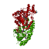
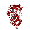
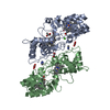
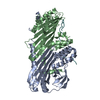
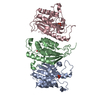
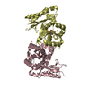
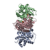
Structure of the azide-inhibited form of cytochrome c peroxidase from obligate human pathogenic bacterium Neisseria gonorrhoeae
Components
Cytochrome-c peroxidase
Keywords
ELECTRON TRANSPORT / Neisseria gonorrhoeae / bacterial peroxidase / ROS detoxification
Function / homology
Function and homology information
cytochrome-c peroxidase / cytochrome-c peroxidase activity / periplasmic space / electron transfer activity / heme binding / metal ion binding Similarity search - Function
Di-c-type haem protein, MauG/cytochrome c peroxidase / Di-haem cytochrome c peroxidase / Di-haem cytochrome c peroxidase / : / Cytochrome c / Cytochrome c-like domain / Cytochrome Bc1 Complex; Chain D, domain 2 / Cytochrome c family profile. / Cytochrome c-like domain / Cytochrome c-like domain superfamily ...Di-c-type haem protein, MauG/cytochrome c peroxidase / Di-haem cytochrome c peroxidase / Di-haem cytochrome c peroxidase / : / Cytochrome c / Cytochrome c-like domain / Cytochrome Bc1 Complex; Chain D, domain 2 / Cytochrome c family profile. / Cytochrome c-like domain / Cytochrome c-like domain superfamily / Prokaryotic membrane lipoprotein lipid attachment site profile. / Orthogonal Bundle / Mainly Alpha Similarity search - Domain/homology
Mass: 18.015 Da / Num. of mol.: 187 / Source method: isolated from a natural source / Formula: H2O
Has ligand of interest
Y
Has protein modification
Y
-
Experimental details
-
Experiment
Experiment
Method: X-RAY DIFFRACTION / Number of used crystals: 1
-
Sample preparation
Crystal
Density Matthews: 2.3 Å3/Da / Density % sol: 46.61 %
Crystal grow
Temperature: 293 K / Method: vapor diffusion Details: 30 % 5/4 PO/OH and 0.1 M MES pH 6.0 in the presence of 2 mM CaCl2, 10 mM sodium ascorbate and 0.2 mM FMN
-
Data collection
Diffraction
Mean temperature: 110 K / Serial crystal experiment: N
Resolution: 2.3→64.95 Å / Cor.coef. Fo:Fc: 0.957 / Cor.coef. Fo:Fc free: 0.929 / SU B: 13.06 / SU ML: 0.168 / Cross valid method: THROUGHOUT / ESU R: 0.353 / ESU R Free: 0.219 / Stereochemistry target values: MAXIMUM LIKELIHOOD Details: U VALUES : WITH TLS ADDED HYDROGENS HAVE BEEN ADDED IN THE RIDING POSITIONS U VALUES : RESIDUAL ONLY
Rfactor
Num. reflection
% reflection
Selection details
Rfree
0.21461
1466
4.9 %
RANDOM
Rwork
0.1712
-
-
-
obs
0.17336
28748
99.18 %
-
Solvent computation
Ion probe radii: 0.7 Å / Shrinkage radii: 0.7 Å / VDW probe radii: 1 Å / Solvent model: MASK
Displacement parameters
Biso mean: 39.118 Å2
Baniso -1
Baniso -2
Baniso -3
1-
0.39 Å2
0 Å2
0 Å2
2-
-
-1.5 Å2
0 Å2
3-
-
-
1.11 Å2
Refinement step
Cycle: LAST / Resolution: 2.3→64.95 Å
Protein
Nucleic acid
Ligand
Solvent
Total
Num. atoms
5059
0
180
187
5426
Refine LS restraints
Refine-ID
Type
Dev ideal
Dev ideal target
Number
X-RAY DIFFRACTION
r_bond_refined_d
0.01
0.018
5410
X-RAY DIFFRACTION
r_bond_other_d
0.001
0.019
4991
X-RAY DIFFRACTION
r_angle_refined_deg
1.907
1.919
7358
X-RAY DIFFRACTION
r_angle_other_deg
1.106
2.737
11518
X-RAY DIFFRACTION
r_dihedral_angle_1_deg
6.05
5
652
X-RAY DIFFRACTION
r_dihedral_angle_2_deg
35.888
23.773
273
X-RAY DIFFRACTION
r_dihedral_angle_3_deg
13.779
15
880
X-RAY DIFFRACTION
r_dihedral_angle_4_deg
15.076
15
26
X-RAY DIFFRACTION
r_chiral_restr
0.108
0.2
752
X-RAY DIFFRACTION
r_gen_planes_refined
0.007
0.02
6224
X-RAY DIFFRACTION
r_gen_planes_other
0.007
0.02
1232
X-RAY DIFFRACTION
r_mcbond_it
1.515
2.011
2620
X-RAY DIFFRACTION
r_mcbond_other
1.501
2.009
2611
X-RAY DIFFRACTION
r_mcangle_it
2.304
3.006
3257
X-RAY DIFFRACTION
r_mcangle_other
2.304
3.007
3258
X-RAY DIFFRACTION
r_scbond_it
2.315
2.262
2790
X-RAY DIFFRACTION
r_scbond_other
2.197
2.26
2786
X-RAY DIFFRACTION
r_scangle_other
3.227
3.311
4099
X-RAY DIFFRACTION
r_long_range_B_refined
4.952
24.292
6288
X-RAY DIFFRACTION
r_long_range_B_other
4.934
24.244
6272
LS refinement shell
Resolution: 2.3→2.36 Å / Total num. of bins used: 20
Rfactor
Num. reflection
% reflection
Rfree
0.26
99
-
Rwork
0.251
2070
-
obs
-
-
98.19 %
Refinement TLS params.
Method: refined / Refine-ID: X-RAY DIFFRACTION
ID
L11 (°2)
L12 (°2)
L13 (°2)
L22 (°2)
L23 (°2)
L33 (°2)
S11 (Å °)
S12 (Å °)
S13 (Å °)
S21 (Å °)
S22 (Å °)
S23 (Å °)
S31 (Å °)
S32 (Å °)
S33 (Å °)
T11 (Å2)
T12 (Å2)
T13 (Å2)
T22 (Å2)
T23 (Å2)
T33 (Å2)
Origin x (Å)
Origin y (Å)
Origin z (Å)
1
2.0828
-1.0535
0.5485
2.4737
-0.3758
1.3062
0.0022
0.1092
-0.0375
-0.1521
0.0034
0.4668
-0.0354
-0.1723
-0.0057
0.0551
-0.0292
-0.0429
0.1327
-0.0026
0.1813
-26.983
-2.478
13.141
2
1.7628
-1.1173
-0.103
3.1114
-0.0242
0.8259
0.0883
0.1119
-0.0073
-0.1836
-0.0613
-0.1866
-0.0139
0.0609
-0.027
0.0558
-0.0212
0.0188
0.0485
-0.034
0.0478
9.354
-4.156
14.715
Refinement TLS group
ID
Refine-ID
Refine TLS-ID
Auth asym-ID
Auth seq-ID
1
X-RAY DIFFRACTION
1
A
1 - 329
2
X-RAY DIFFRACTION
2
B
3 - 325
+
About Yorodumi
-
News
-
Feb 9, 2022. New format data for meta-information of EMDB entries
New format data for meta-information of EMDB entries
Version 3 of the EMDB header file is now the official format.
The previous official version 1.9 will be removed from the archive.
In the structure databanks used in Yorodumi, some data are registered as the other names, "COVID-19 virus" and "2019-nCoV". Here are the details of the virus and the list of structure data.
Jan 31, 2019. EMDB accession codes are about to change! (news from PDBe EMDB page)
EMDB accession codes are about to change! (news from PDBe EMDB page)
The allocation of 4 digits for EMDB accession codes will soon come to an end. Whilst these codes will remain in use, new EMDB accession codes will include an additional digit and will expand incrementally as the available range of codes is exhausted. The current 4-digit format prefixed with “EMD-” (i.e. EMD-XXXX) will advance to a 5-digit format (i.e. EMD-XXXXX), and so on. It is currently estimated that the 4-digit codes will be depleted around Spring 2019, at which point the 5-digit format will come into force.
The EM Navigator/Yorodumi systems omit the EMD- prefix.
Related info.:Q: What is EMD? / ID/Accession-code notation in Yorodumi/EM Navigator
Yorodumi is a browser for structure data from EMDB, PDB, SASBDB, etc.
This page is also the successor to EM Navigator detail page, and also detail information page/front-end page for Omokage search.
The word "yorodu" (or yorozu) is an old Japanese word meaning "ten thousand". "mi" (miru) is to see.
Related info.:EMDB / PDB / SASBDB / Comparison of 3 databanks / Yorodumi Search / Aug 31, 2016. New EM Navigator & Yorodumi / Yorodumi Papers / Jmol/JSmol / Function and homology information / Changes in new EM Navigator and Yorodumi
 Movie
Movie Controller

 Yorodumi
 Open data
 Basic information
 Components
 Keywords
 Function and homology information
 Neisseria gonorrhoeae (bacteria)
 X-RAY DIFFRACTION /
  Authors
 Portugal, 2items
  Citation
 Structure visualization
 Downloads & links
 Other downloads
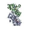


 PDBj
PDBj


 Assembly
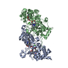

 Mass: 618.503 Da / Num. of mol.: 4 / Source method: obtained synthetically / Formula: C34H34FeN4O4 / Feature type: SUBJECT OF INVESTIGATION
 Mass: 40.078 Da / Num. of mol.: 2 / Source method: obtained synthetically / Formula: Ca
 Mass: 42.020 Da / Num. of mol.: 2 / Source method: obtained synthetically / Formula: N3
 Mass: 18.015 Da / Num. of mol.: 187 / Source method: isolated from a natural source / Formula: H2O
 Sample preparation
 / Beamline: BM30A / Wavelength: 0.979 Å
 Processing