 Movie
Movie Controller

[English] 日本語
 Yorodumi
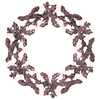
- PDB-5muf: Crystal structure of human phosphoglycerate mutase family member ... -
+ Open data

- Basic information
| Entry | Database: PDB / ID: 5muf | ||||||
|---|---|---|---|---|---|---|---|
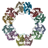
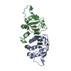
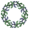
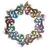
| Title | Crystal structure of human phosphoglycerate mutase family member 5 (PGAM5) in its enzymatically active dodecameric form induced by the presence of the N-terminal WDPNWD motif | ||||||
 Components Components | Serine/threonine-protein phosphatase PGAM5, mitochondrial | ||||||
 Keywords Keywords | HYDROLASE / PHOSPHOGLYCERATE MUTASE FAMILY MEMBER 5 / PGAM5 / SERINE/THREONINE PHOSPHATASE / MITOCHONDRIAL PROTEIN / WDPNWD motif / WDXNWD motif / dimer / dodecamer / Structural Genomics / Structural Genomics Consortium / SGC | ||||||
| Function / homology |  Function and homology information Function and homology informationnegative regulation of cold-induced thermogenesis / positive regulation of mitochondrial fission / necroptotic process / protein-serine/threonine phosphatase / Receptor Mediated Mitophagy / protein serine/threonine phosphatase activity / Regulation of pyruvate metabolism / macroautophagy / mitochondrial outer membrane / mitochondrial inner membrane / mitochondrion Similarity search - Function | ||||||
| Biological species |  Homo sapiens (human) Homo sapiens (human) | ||||||
| Method |  X-RAY DIFFRACTION / SYNCHROTRON / MOLECULAR REPLACEMENT / Resolution: 3.1 Å X-RAY DIFFRACTION / SYNCHROTRON / MOLECULAR REPLACEMENT / Resolution: 3.1 Å | ||||||
 Authors Authors | Chaikuad, A. / Alfano, I. / Picaud, S. / Filippakopoulos, P. / von Delft, F. / Bountra, C. / Arrowsmith, C.H. / Edwards, A.M. / Knapp, S. / Structural Genomics Consortium (SGC) | ||||||
 Citation Citation | Journal: Structure / Year: 2017 Title: Structures of PGAM5 Provide Insight into Active Site Plasticity and Multimeric Assembly. Authors: Chaikuad, A. / Filippakopoulos, P. / Marcsisin, S.R. / Picaud, S. / Schroder, M. / Sekine, S. / Ichijo, H. / Engen, J.R. / Takeda, K. / Knapp, S. | ||||||
| History |
|
- Structure visualization
| Structure viewer | Molecule: MolmilJmol/JSmol |
|---|
- Downloads & links
-Download
| PDBx/mmCIF format | 5muf.cif.gz | 258.3 KB | Display | PDBx/mmCIF format |
|---|---|---|---|---|
| PDB format | pdb5muf.ent.gz | 210.3 KB | Display | PDB format |
| PDBx/mmJSON format | 5muf.json.gz | Tree view | PDBx/mmJSON format | |
| Others |  Other downloads Other downloads |
-Validation report
| Arichive directory | https://data.pdbj.org/pub/pdb/validation_reports/mu/5mufftp://data.pdbj.org/pub/pdb/validation_reports/mu/5muf | HTTPS FTP |
|---|
-Related structure data
| Related structure data | 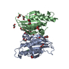 3mxoSC  3o0tC C: citing same article ( S: Starting model for refinement |
|---|---|
| Similar structure data |

-Links
 PDBj
PDBj


- Assembly
| Deposited unit | 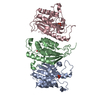
| |||||||||||||||||||||||||||||||||||||||||||||||||||||
|---|---|---|---|---|---|---|---|---|---|---|---|---|---|---|---|---|---|---|---|---|---|---|---|---|---|---|---|---|---|---|---|---|---|---|---|---|---|---|---|---|---|---|---|---|---|---|---|---|---|---|---|---|---|---|
| 1 | 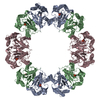
| |||||||||||||||||||||||||||||||||||||||||||||||||||||
| Unit cell |
| |||||||||||||||||||||||||||||||||||||||||||||||||||||
| Noncrystallographic symmetry (NCS) | NCS domain:
NCS domain segments: Component-ID: _ / Beg auth comp-ID: GLY / Beg label comp-ID: GLY / End auth comp-ID: SER / End label comp-ID: SER / Refine code: _ / Auth seq-ID: 56 - 289 / Label seq-ID: 5 - 238
NCS ensembles :
|
-Components
| #1: Protein | Mass: 27138.895 Da / Num. of mol.: 3 Source method: isolated from a genetically manipulated source Source: (gene. exp.) Homo sapiens (human) / Gene: PGAM5 / Plasmid: pNIC28-Bsa4 / Production host:  References: UniProt: Q96HS1, protein-serine/threonine phosphatase #2: Chemical |   Mass: 94.971 Da / Num. of mol.: 3 / Source method: obtained synthetically / Formula: PO4 Mass: 94.971 Da / Num. of mol.: 3 / Source method: obtained synthetically / Formula: PO4#3: Water | ChemComp-HOH / |  Mass: 18.015 Da / Num. of mol.: 51 / Source method: isolated from a natural source / Formula: H2O Mass: 18.015 Da / Num. of mol.: 51 / Source method: isolated from a natural source / Formula: H2O |
|---|
-Experimental details
-Experiment
| Experiment | Method: X-RAY DIFFRACTION / Number of used crystals: 1 |
|---|
- Sample preparation
| Crystal | Density Matthews: 3.27 Å3/Da / Density % sol: 62.37 % |
|---|---|
| Crystal grow | Temperature: 293 K / Method: vapor diffusion, sitting drop / pH: 5.7 / Details: 18% PEG 3350 and 0.1 M MES, pH 5.7 |
-Data collection
| Diffraction | Mean temperature: 100 K |
|---|---|
| Diffraction source | Source: SYNCHROTRON / Site: Diamond  / Beamline: I03 / Wavelength: 0.9763 Å / Beamline: I03 / Wavelength: 0.9763 Å |
| Detector | Type: DECTRIS PILATUS3 6M / Detector: PIXEL / Date: Apr 25, 2011 |
| Radiation | Protocol: SINGLE WAVELENGTH / Monochromatic (M) / Laue (L): M / Scattering type: x-ray |
| Radiation wavelength | Wavelength: 0.9763 Å / Relative weight: 1 |
| Reflection | Resolution: 3.1→41.03 Å / Num. obs: 18243 / % possible obs: 94.3 % / Redundancy: 7 % / Biso Wilson estimate: 55.3 Å2 / Rmerge(I) obs: 0.271 / Rpim(I) all: 0.101 / Net I/σ(I): 6.8 |
| Reflection shell | Resolution: 3.1→3.27 Å / Redundancy: 6.9 % / Rmerge(I) obs: 0.993 / Mean I/σ(I) obs: 2 / Num. unique obs: 2709 / Rpim(I) all: 0.373 / % possible all: 96.2 |
- Processing
| Software |
| ||||||||||||||||||||||||||||||||||||||||||||||||||||||||||||||||||||||||||||||||||||||||||||||||||||||||||||||||||||||||||||||||||||||||||||||||||||||||||||||||||||||||||||||||||||||
|---|---|---|---|---|---|---|---|---|---|---|---|---|---|---|---|---|---|---|---|---|---|---|---|---|---|---|---|---|---|---|---|---|---|---|---|---|---|---|---|---|---|---|---|---|---|---|---|---|---|---|---|---|---|---|---|---|---|---|---|---|---|---|---|---|---|---|---|---|---|---|---|---|---|---|---|---|---|---|---|---|---|---|---|---|---|---|---|---|---|---|---|---|---|---|---|---|---|---|---|---|---|---|---|---|---|---|---|---|---|---|---|---|---|---|---|---|---|---|---|---|---|---|---|---|---|---|---|---|---|---|---|---|---|---|---|---|---|---|---|---|---|---|---|---|---|---|---|---|---|---|---|---|---|---|---|---|---|---|---|---|---|---|---|---|---|---|---|---|---|---|---|---|---|---|---|---|---|---|---|---|---|---|---|
| Refinement | Method to determine structure: MOLECULAR REPLACEMENT Starting model: 3MXO Resolution: 3.1→41.03 Å / Cor.coef. Fo:Fc: 0.904 / Cor.coef. Fo:Fc free: 0.868 / SU B: 46.834 / SU ML: 0.369 / Cross valid method: THROUGHOUT / ESU R Free: 0.451 / Details: HYDROGENS HAVE BEEN ADDED IN THE RIDING POSITIONS
| ||||||||||||||||||||||||||||||||||||||||||||||||||||||||||||||||||||||||||||||||||||||||||||||||||||||||||||||||||||||||||||||||||||||||||||||||||||||||||||||||||||||||||||||||||||||
| Solvent computation | Ion probe radii: 0.8 Å / Shrinkage radii: 0.8 Å / VDW probe radii: 1.2 Å | ||||||||||||||||||||||||||||||||||||||||||||||||||||||||||||||||||||||||||||||||||||||||||||||||||||||||||||||||||||||||||||||||||||||||||||||||||||||||||||||||||||||||||||||||||||||
| Displacement parameters | Biso mean: 50.597 Å2
| ||||||||||||||||||||||||||||||||||||||||||||||||||||||||||||||||||||||||||||||||||||||||||||||||||||||||||||||||||||||||||||||||||||||||||||||||||||||||||||||||||||||||||||||||||||||
| Refinement step | Cycle: 1 / Resolution: 3.1→41.03 Å
| ||||||||||||||||||||||||||||||||||||||||||||||||||||||||||||||||||||||||||||||||||||||||||||||||||||||||||||||||||||||||||||||||||||||||||||||||||||||||||||||||||||||||||||||||||||||
| Refine LS restraints |
|