 Movie
Movie Controller

[English] 日本語
 Yorodumi
- PDB-6fu3: Structure of the mixed-valence, active form, of cytochrome c pero... -
+ Open data

- Basic information
| Entry | Database: PDB / ID: 6fu3 | |||||||||
|---|---|---|---|---|---|---|---|---|---|---|
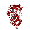
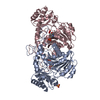
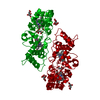
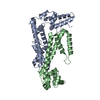
| Title | Structure of the mixed-valence, active form, of cytochrome c peroxidase from obligate human pathogenic bacterium Neisseria gonorrhoeae | |||||||||
 Components Components | Protein CcpR | |||||||||
 Keywords Keywords | ELECTRON TRANSPORT / Neisseria gonorrhoeae / bacterial peroxidase / ROS detoxification | |||||||||
| Function / homology |  Function and homology information Function and homology informationcytochrome-c peroxidase / cytochrome-c peroxidase activity / electron transfer activity / periplasmic space / heme binding / metal ion binding Similarity search - Function | |||||||||
| Biological species |  Neisseria gonorrhoeae (bacteria) Neisseria gonorrhoeae (bacteria) | |||||||||
| Method |  X-RAY DIFFRACTION / MOLECULAR REPLACEMENT / Resolution: 1.8 Å X-RAY DIFFRACTION / MOLECULAR REPLACEMENT / Resolution: 1.8 Å | |||||||||
 Authors Authors | Carvalho, A.L. / Romao, M.J. / Pauleta, S. / Nobrega, C. | |||||||||
| Funding support |  Portugal, 2items Portugal, 2items
| |||||||||
 Citation Citation | Journal: To Be Published Title: Structure of the mixed-valence, active form, of cytochrome c peroxidase from obligate human pathogenic bacterium Neisseria gonorrhoeae Authors: Carvalho, A.L. / Romao, M.J. / Pauleta, S. / Nobrega, C. | |||||||||
| History |
|
- Structure visualization
| Structure viewer | Molecule: MolmilJmol/JSmol |
|---|
- Downloads & links
-Download
| PDBx/mmCIF format | 6fu3.cif.gz | 278.8 KB | Display | PDBx/mmCIF format |
|---|---|---|---|---|
| PDB format | pdb6fu3.ent.gz | 224.8 KB | Display | PDB format |
| PDBx/mmJSON format | 6fu3.json.gz | Tree view | PDBx/mmJSON format | |
| Others |  Other downloads Other downloads |
-Validation report
| Arichive directory | https://data.pdbj.org/pub/pdb/validation_reports/fu/6fu3ftp://data.pdbj.org/pub/pdb/validation_reports/fu/6fu3 | HTTPS FTP |
|---|
-Related structure data
| Related structure data |  6qknC  2vhdS S: Starting model for refinement C: citing same article ( |
|---|---|
| Similar structure data |


-Links
 PDBj
PDBj


- Assembly
| Deposited unit | 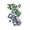
| ||||||||
|---|---|---|---|---|---|---|---|---|---|
| 1 |
| ||||||||
| Unit cell |
|
-Components
| #1: Protein | Mass: 37617.297 Da / Num. of mol.: 2 Source method: isolated from a genetically manipulated source Source: (gene. exp.) Neisseria gonorrhoeae (bacteria)Gene: ccpA, WHOF_00612, WHOF_02054, WHOG_00329, WHOG_02247C, WHOK_00254, WHOK_02247C, WHOL_00503, WHOL_01944C, WHOM_00704, WHOM_02256C, WHON_00634, WHON_02251C, WHOO_00344, WHOO_01926, WHOP_00646, ...Gene: ccpA, WHOF_00612, WHOF_02054, WHOG_00329, WHOG_02247C, WHOK_00254, WHOK_02247C, WHOL_00503, WHOL_01944C, WHOM_00704, WHOM_02256C, WHON_00634, WHON_02251C, WHOO_00344, WHOO_01926, WHOP_00646, WHOP_02256C, WHOU_00764, WHOU_02329C, WHOV_00550, WHOV_02317C, WHOW_00232, WHOW_02310C, WHOX_00887, WHOX_02246C, WHOY_00586, WHOY_02329C, WHOZ_00270, WHOZ_02318C Production host: References: UniProt: A0A1D3HIT0, UniProt: Q5F5Z9*PLUS, cytochrome-c peroxidase #2: Chemical |   Mass: 40.078 Da / Num. of mol.: 2 / Source method: obtained synthetically / Formula: Ca Mass: 40.078 Da / Num. of mol.: 2 / Source method: obtained synthetically / Formula: Ca#3: Chemical | ChemComp-HEC /   Mass: 618.503 Da / Num. of mol.: 4 / Source method: obtained synthetically / Formula: C34H34FeN4O4 / Feature type: SUBJECT OF INVESTIGATION Mass: 618.503 Da / Num. of mol.: 4 / Source method: obtained synthetically / Formula: C34H34FeN4O4 / Feature type: SUBJECT OF INVESTIGATION#4: Water | ChemComp-HOH / |  Mass: 18.015 Da / Num. of mol.: 524 / Source method: isolated from a natural source / Formula: H2O Mass: 18.015 Da / Num. of mol.: 524 / Source method: isolated from a natural source / Formula: H2OHas ligand of interest | Y | Has protein modification | Y | |
|---|
-Experimental details
-Experiment
| Experiment | Method: X-RAY DIFFRACTION / Number of used crystals: 1 |
|---|
- Sample preparation
| Crystal | Density Matthews: 2.27 Å3/Da / Density % sol: 45.71 % |
|---|---|
| Crystal grow | Temperature: 278 K / Method: vapor diffusion / pH: 7.5 Details: 30% 5/4 PO/OH and 0.1M MES pH6.0 in the presence of 2mM CaCl2, 10mM sodium ascorbate and 0.2mM FMN, using a 20mg/mL protein solution previously incubated with calcium, sodium ascorbate and FMN. |
-Data collection
| Diffraction | Mean temperature: 110 K / Serial crystal experiment: N |
|---|---|
| Diffraction source | Source: SEALED TUBE / Type: BRUKER IMUS MICROFOCUS / Wavelength: 1.5418 Å |
| Detector | Type: BRUKER PHOTON 100 / Detector: CMOS / Date: Apr 4, 2016 |
| Radiation | Monochromator: Ni filter / Protocol: SINGLE WAVELENGTH / Monochromatic (M) / Laue (L): M / Scattering type: x-ray |
| Radiation wavelength | Wavelength: 1.5418 Å / Relative weight: 1 |
| Reflection | Resolution: 1.79→64.3 Å / Num. obs: 61589 / % possible obs: 99.1 % / Redundancy: 9.2 % / Rmerge(I) obs: 0.1565 / Net I/σ(I): 10.3 |
| Reflection shell | Resolution: 1.79→1.82 Å / Rmerge(I) obs: 0.8398 / Num. unique obs: 2299 |
- Processing
| Software |
| |||||||||||||||||||||||||||||||||||||||||||||||||||||||||||||||||||||||||||||||||||||||||||||||||||||||||
|---|---|---|---|---|---|---|---|---|---|---|---|---|---|---|---|---|---|---|---|---|---|---|---|---|---|---|---|---|---|---|---|---|---|---|---|---|---|---|---|---|---|---|---|---|---|---|---|---|---|---|---|---|---|---|---|---|---|---|---|---|---|---|---|---|---|---|---|---|---|---|---|---|---|---|---|---|---|---|---|---|---|---|---|---|---|---|---|---|---|---|---|---|---|---|---|---|---|---|---|---|---|---|---|---|---|---|
| Refinement | Method to determine structure: MOLECULAR REPLACEMENT Starting model: 2VHD Resolution: 1.8→23.82 Å / Cor.coef. Fo:Fc: 0.949 / Cor.coef. Fo:Fc free: 0.928 / SU B: 6.836 / SU ML: 0.114 / Cross valid method: THROUGHOUT / ESU R: 0.156 / ESU R Free: 0.142 / Stereochemistry target values: MAXIMUM LIKELIHOOD Details: U VALUES : WITH TLS ADDED HYDROGENS HAVE BEEN ADDED IN THE RIDING POSITIONS U VALUES : RESIDUAL ONLY
| |||||||||||||||||||||||||||||||||||||||||||||||||||||||||||||||||||||||||||||||||||||||||||||||||||||||||
| Solvent computation | Ion probe radii: 0.8 Å / Shrinkage radii: 0.8 Å / VDW probe radii: 1.3 Å / Solvent model: MASK | |||||||||||||||||||||||||||||||||||||||||||||||||||||||||||||||||||||||||||||||||||||||||||||||||||||||||
| Displacement parameters | Biso mean: 21.856 Å2
| |||||||||||||||||||||||||||||||||||||||||||||||||||||||||||||||||||||||||||||||||||||||||||||||||||||||||
| Refinement step | Cycle: LAST / Resolution: 1.8→23.82 Å
| |||||||||||||||||||||||||||||||||||||||||||||||||||||||||||||||||||||||||||||||||||||||||||||||||||||||||
| Refine LS restraints |
| |||||||||||||||||||||||||||||||||||||||||||||||||||||||||||||||||||||||||||||||||||||||||||||||||||||||||
| LS refinement shell | Resolution: 1.8→1.847 Å / Total num. of bins used: 20
| |||||||||||||||||||||||||||||||||||||||||||||||||||||||||||||||||||||||||||||||||||||||||||||||||||||||||
| Refinement TLS params. | Method: refined / Refine-ID: X-RAY DIFFRACTION
| |||||||||||||||||||||||||||||||||||||||||||||||||||||||||||||||||||||||||||||||||||||||||||||||||||||||||
| Refinement TLS group |
|
