 Movie
Movie Controller

[English] 日本語
 Yorodumi
- PDB-1ibj: Crystal structure of cystathionine beta-lyase from Arabidopsis th... -
+ Open data

- Basic information
| Entry | Database: PDB / ID: 1ibj | ||||||
|---|---|---|---|---|---|---|---|
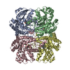
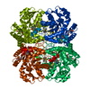
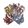
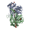
| Title | Crystal structure of cystathionine beta-lyase from Arabidopsis thaliana | ||||||
 Components Components | CYSTATHIONINE BETA-LYASE | ||||||
 Keywords Keywords | LYASE / PLP-dependent enzyme / Methionine biosynthesis / Transsulfuration | ||||||
| Function / homology |  Function and homology information Function and homology information: / carbon-sulfur lyase activity / cysteine-S-conjugate beta-lyase activity / cysteine-S-conjugate beta-lyase / L-methionine metabolic process / transsulfuration / chloroplast stroma / chloroplast / pyridoxal phosphate binding / molecular adaptor activity ...: / carbon-sulfur lyase activity / cysteine-S-conjugate beta-lyase activity / cysteine-S-conjugate beta-lyase / L-methionine metabolic process / transsulfuration / chloroplast stroma / chloroplast / pyridoxal phosphate binding / molecular adaptor activity / protein homodimerization activity / cytoplasm Similarity search - Function | ||||||
| Biological species |  | ||||||
| Method |  X-RAY DIFFRACTION / SYNCHROTRON / MOLECULAR REPLACEMENT / Resolution: 2.3 Å X-RAY DIFFRACTION / SYNCHROTRON / MOLECULAR REPLACEMENT / Resolution: 2.3 Å | ||||||
 Authors Authors | Breitinger, U. / Clausen, T. / Messerschmidt, A. | ||||||
 Citation Citation | Journal: Plant Physiol. / Year: 2001 Title: The three-dimensional structure of cystathionine beta-lyase from Arabidopsis and its substrate specificity Authors: Breitinger, U. / Clausen, T. / Ehlert, S. / Huber, R. / Laber, B. / Schmidt, F. / Pohl, E. / Messerschmidt, A. | ||||||
| History |
|
- Structure visualization
| Structure viewer | Molecule: MolmilJmol/JSmol |
|---|
- Downloads & links
-Download
| PDBx/mmCIF format | 1ibj.cif.gz | 161.7 KB | Display | PDBx/mmCIF format |
|---|---|---|---|---|
| PDB format | pdb1ibj.ent.gz | 126.2 KB | Display | PDB format |
| PDBx/mmJSON format | 1ibj.json.gz | Tree view | PDBx/mmJSON format | |
| Others |  Other downloads Other downloads |
-Validation report
| Arichive directory | https://data.pdbj.org/pub/pdb/validation_reports/ib/1ibjftp://data.pdbj.org/pub/pdb/validation_reports/ib/1ibj | HTTPS FTP |
|---|
-Related structure data
| Related structure data |  1qgnS S: Starting model for refinement |
|---|---|
| Similar structure data |


-Links
 PDBj
PDBj


- Assembly
| Deposited unit | 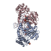
| ||||||||
|---|---|---|---|---|---|---|---|---|---|
| 1 | 
| ||||||||
| Unit cell |
| ||||||||
| Details | Two active dimers, related by 2-fold crystallographic symmetry, form the homotetramer. |
-Components
| #1: Protein | Mass: 50490.480 Da / Num. of mol.: 2 Source method: isolated from a genetically manipulated source Source: (gene. exp.)  #2: Chemical |   Mass: 60.009 Da / Num. of mol.: 2 / Source method: obtained synthetically / Formula: CO3 Mass: 60.009 Da / Num. of mol.: 2 / Source method: obtained synthetically / Formula: CO3#3: Chemical | ChemComp-SO4 / |   Mass: 96.063 Da / Num. of mol.: 1 / Source method: obtained synthetically / Formula: SO4 Mass: 96.063 Da / Num. of mol.: 1 / Source method: obtained synthetically / Formula: SO4#4: Chemical |   Mass: 247.142 Da / Num. of mol.: 2 / Source method: obtained synthetically / Formula: C8H10NO6P Mass: 247.142 Da / Num. of mol.: 2 / Source method: obtained synthetically / Formula: C8H10NO6P#5: Water | ChemComp-HOH / |  Mass: 18.015 Da / Num. of mol.: 253 / Source method: isolated from a natural source / Formula: H2O Mass: 18.015 Da / Num. of mol.: 253 / Source method: isolated from a natural source / Formula: H2O |
|---|
-Experimental details
-Experiment
| Experiment | Method: X-RAY DIFFRACTION / Number of used crystals: 1 |
|---|
- Sample preparation
| Crystal | Density Matthews: 2.44 Å3/Da / Density % sol: 49.69 % | ||||||||||||||||||||||||||||||||||||||||||
|---|---|---|---|---|---|---|---|---|---|---|---|---|---|---|---|---|---|---|---|---|---|---|---|---|---|---|---|---|---|---|---|---|---|---|---|---|---|---|---|---|---|---|---|
| Crystal grow | Temperature: 294 K / Method: vapor diffusion, sitting drop / pH: 6 Details: Ammonium sulfate, Mes-NaOH, PLP, pH 6.0, VAPOR DIFFUSION, SITTING DROP, temperature 294K | ||||||||||||||||||||||||||||||||||||||||||
| Crystal grow | *PLUS | ||||||||||||||||||||||||||||||||||||||||||
| Components of the solutions | *PLUS
|
-Data collection
| Diffraction | Mean temperature: 100 K |
|---|---|
| Diffraction source | Source: SYNCHROTRON / Site: EMBL/DESY, HAMBURG  / Beamline: BW7B / Wavelength: 0.83 Å / Beamline: BW7B / Wavelength: 0.83 Å |
| Detector | Type: MARRESEARCH / Detector: IMAGE PLATE / Date: Oct 25, 1999 |
| Radiation | Monochromator: Si 111 Channel / Protocol: SINGLE WAVELENGTH / Monochromatic (M) / Laue (L): M / Scattering type: x-ray |
| Radiation wavelength | Wavelength: 0.83 Å / Relative weight: 1 |
| Reflection | Resolution: 2.3→15 Å / Num. all: 108362 / Num. obs: 102185 / % possible obs: 94.3 % / Observed criterion σ(F): 2 / Observed criterion σ(I): 2 / Redundancy: 3.8 % / Rmerge(I) obs: 0.068 / Rsym value: 0.072 / Net I/σ(I): 22.3 |
| Reflection shell | Resolution: 2.3→2.38 Å / Redundancy: 2.1 % / Rmerge(I) obs: 0.135 / Mean I/σ(I) obs: 3.1 / Num. unique all: 39111 / Rsym value: 0.141 / % possible all: 81.6 |
| Reflection | *PLUS Num. obs: 37727 / Num. measured all: 102185 |
| Reflection shell | *PLUS % possible obs: 81.6 % |
- Processing
| Software |
| |||||||||||||||||||||||||
|---|---|---|---|---|---|---|---|---|---|---|---|---|---|---|---|---|---|---|---|---|---|---|---|---|---|---|
| Refinement | Method to determine structure: MOLECULAR REPLACEMENT Starting model: PDB ENTRY 1QGN Resolution: 2.3→6 Å / Isotropic thermal model: Anisotropic / Cross valid method: THROUGHOUT / σ(F): 2 / σ(I): 2 / Stereochemistry target values: Engh & Huber
| |||||||||||||||||||||||||
| Displacement parameters |
| |||||||||||||||||||||||||
| Refinement step | Cycle: LAST / Resolution: 2.3→6 Å
| |||||||||||||||||||||||||
| Refine LS restraints |
| |||||||||||||||||||||||||
| Software | *PLUS Name: CNS / Version: 1 / Classification: refinement | |||||||||||||||||||||||||
| Refinement | *PLUS Rfactor Rfree: 0.309 / Rfactor Rwork: 0.249 | |||||||||||||||||||||||||
| Solvent computation | *PLUS | |||||||||||||||||||||||||
| Displacement parameters | *PLUS |
