 Movie
Movie Controller

[English] 日本語
 Yorodumi
- PDB-1cl2: CYSTATHIONINE BETA-LYASE (CBL) FROM ESCHERICHIA COLI IN COMPLEX W... -
+ Open data

- Basic information
| Entry | Database: PDB / ID: 1cl2 | |||||||||
|---|---|---|---|---|---|---|---|---|---|---|
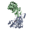
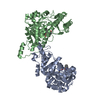
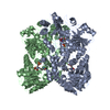
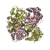
| Title | CYSTATHIONINE BETA-LYASE (CBL) FROM ESCHERICHIA COLI IN COMPLEX WITH AMINOETHOXYVINYLGLYCINE | |||||||||
 Components Components | CYSTATHIONINE BETA-LYASE | |||||||||
 Keywords Keywords | METHIONINE BIOSYNTHESIS / PLP-DEPENDENT ENZYMES / C-S BETA LYASE / AMINOETHOXYVINYLGLYCINE / SLOW-BINDING INHIBITION | |||||||||
| Function / homology |  Function and homology information Function and homology informationL-cysteine desulfidase / : / cysteine-S-conjugate beta-lyase activity / cysteine-S-conjugate beta-lyase / alanine racemase activity / L-cysteine desulfhydrase activity / transsulfuration / : / pyridoxal phosphate binding / protein homotetramerization ...L-cysteine desulfidase / : / cysteine-S-conjugate beta-lyase activity / cysteine-S-conjugate beta-lyase / alanine racemase activity / L-cysteine desulfhydrase activity / transsulfuration / : / pyridoxal phosphate binding / protein homotetramerization / protein-containing complex / identical protein binding / cytoplasm Similarity search - Function | |||||||||
| Biological species |  | |||||||||
| Method |  X-RAY DIFFRACTION / Resolution: 2.2 Å X-RAY DIFFRACTION / Resolution: 2.2 Å | |||||||||
 Authors Authors | Clausen, T. / Huber, R. / Messerschmidt, A. | |||||||||
 Citation Citation | Journal: Biochemistry / Year: 1997 Title: Slow-binding inhibition of Escherichia coli cystathionine beta-lyase by L-aminoethoxyvinylglycine: a kinetic and X-ray study. Authors: Clausen, T. / Huber, R. / Messerschmidt, A. / Pohlenz, H.D. / Laber, B. #1: Journal: J.Mol.Biol. / Year: 1996Title: Crystal Structure of the Pyridoxal-5'-Phosphate Dependent Cystathionine Beta-Lyase from Escherichia Coli at 1.83 A Authors: Clausen, T. / Huber, R. / Laber, B. / Pohlenz, H.D. / Messerschmidt, A. | |||||||||
| History |
|
- Structure visualization
| Structure viewer | Molecule: MolmilJmol/JSmol |
|---|
- Downloads & links
-Download
| PDBx/mmCIF format | 1cl2.cif.gz | 209.7 KB | Display | PDBx/mmCIF format |
|---|---|---|---|---|
| PDB format | pdb1cl2.ent.gz | 169.5 KB | Display | PDB format |
| PDBx/mmJSON format | 1cl2.json.gz | Tree view | PDBx/mmJSON format | |
| Others |  Other downloads Other downloads |
-Validation report
| Arichive directory | https://data.pdbj.org/pub/pdb/validation_reports/cl/1cl2ftp://data.pdbj.org/pub/pdb/validation_reports/cl/1cl2 | HTTPS FTP |
|---|
-Related structure data
| Similar structure data |
|---|


-Links
 PDBj
PDBj- Assembly
| Deposited unit | 
| ||||||||
|---|---|---|---|---|---|---|---|---|---|
| 1 | 
| ||||||||
| Unit cell |
| ||||||||
| Components on special symmetry positions |
| ||||||||
| Noncrystallographic symmetry (NCS) | NCS oper: (Code: given Matrix: (0.997, 0.001, -0.125), Vector: |
-Components
| #1: Protein | Mass: 43261.270 Da / Num. of mol.: 2 Source method: isolated from a genetically manipulated source Source: (gene. exp.) #2: Chemical |   Mass: 389.298 Da / Num. of mol.: 2 / Source method: obtained synthetically / Formula: C14H20N3O8P Mass: 389.298 Da / Num. of mol.: 2 / Source method: obtained synthetically / Formula: C14H20N3O8P#3: Water | ChemComp-HOH / |  Mass: 18.015 Da / Num. of mol.: 430 / Source method: isolated from a natural source / Formula: H2O Mass: 18.015 Da / Num. of mol.: 430 / Source method: isolated from a natural source / Formula: H2O |
|---|
-Experimental details
-Experiment
| Experiment | Method: X-RAY DIFFRACTION / Number of used crystals: 1 |
|---|
- Sample preparation
| Crystal | Density Matthews: 2.1 Å3/Da / Density % sol: 40.84 % | ||||||||||||||||||||
|---|---|---|---|---|---|---|---|---|---|---|---|---|---|---|---|---|---|---|---|---|---|
| Crystal grow | pH: 8.2 / Details: 20% PEG400, 0.15M CACL2, 0.1 M HEPES/NAOH (PH 8.2) | ||||||||||||||||||||
| Crystal grow | *PLUS Method: vapor diffusion, hanging drop / Details: Laber, B., (1996) FEBS Lett., 379, 94. | ||||||||||||||||||||
| Components of the solutions | *PLUS
|
-Data collection
| Diffraction | Mean temperature: 298 K |
|---|---|
| Diffraction source | Source: ROTATING ANODE / Type: RIGAKU RUH2R / Wavelength: 1.5418 |
| Detector | Type: MARRESEARCH / Detector: IMAGE PLATE / Date: Nov 15, 1996 |
| Radiation | Monochromatic (M) / Laue (L): M / Scattering type: x-ray |
| Radiation wavelength | Wavelength: 1.5418 Å / Relative weight: 1 |
| Reflection | Resolution: 2.2→15 Å / % possible obs: 98.1 % / Observed criterion σ(I): 2 / Rmerge(I) obs: 0.065 / Rsym value: 0.063 |
| Reflection shell | Resolution: 2.2→2.3 Å / Mean I/σ(I) obs: 3.5 / % possible all: 94.9 |
- Processing
| Software |
| ||||||||||||||||||||||||||||||||||||||||||||||||||||||||||||
|---|---|---|---|---|---|---|---|---|---|---|---|---|---|---|---|---|---|---|---|---|---|---|---|---|---|---|---|---|---|---|---|---|---|---|---|---|---|---|---|---|---|---|---|---|---|---|---|---|---|---|---|---|---|---|---|---|---|---|---|---|---|
| Refinement | Resolution: 2.2→8 Å / Data cutoff high absF: 1000000 / Data cutoff low absF: 0.001 / σ(F): 2
| ||||||||||||||||||||||||||||||||||||||||||||||||||||||||||||
| Displacement parameters | Biso mean: 16.9 Å2 | ||||||||||||||||||||||||||||||||||||||||||||||||||||||||||||
| Refine analyze | Luzzati coordinate error obs: 0.19 Å | ||||||||||||||||||||||||||||||||||||||||||||||||||||||||||||
| Refinement step | Cycle: LAST / Resolution: 2.2→8 Å
| ||||||||||||||||||||||||||||||||||||||||||||||||||||||||||||
| Refine LS restraints |
| ||||||||||||||||||||||||||||||||||||||||||||||||||||||||||||
| Xplor file |
| ||||||||||||||||||||||||||||||||||||||||||||||||||||||||||||
| Software | *PLUS Name: X-PLOR / Version: 3.1 / Classification: refinement | ||||||||||||||||||||||||||||||||||||||||||||||||||||||||||||
| Refinement | *PLUS Rfactor obs: 0.164 | ||||||||||||||||||||||||||||||||||||||||||||||||||||||||||||
| Solvent computation | *PLUS | ||||||||||||||||||||||||||||||||||||||||||||||||||||||||||||
| Displacement parameters | *PLUS |
