 Movie
Movie Controller

[English] 日本語
 Yorodumi
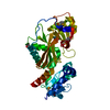
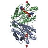
- PDB-1nml: Di-haemic Cytochrome c Peroxidase from Pseudomonas nautica 617, f... -
+ Open data

- Basic information
| Entry | Database: PDB / ID: 1nml | |||||||||
|---|---|---|---|---|---|---|---|---|---|---|
| Title | Di-haemic Cytochrome c Peroxidase from Pseudomonas nautica 617, form IN (pH 4.0) | |||||||||
 Components Components | di-haem cytochrome c peroxidase | |||||||||
 Keywords Keywords | OXIDOREDUCTASE / PEROXIDASE / DI-HAEM / ELECTRON TRANSPORT | |||||||||
| Function / homology |  Function and homology information Function and homology informationcytochrome-c peroxidase activity / electron transfer activity / periplasmic space / heme binding / metal ion binding Similarity search - Function | |||||||||
| Biological species |  Marinobacter hydrocarbonoclasticus (bacteria) Marinobacter hydrocarbonoclasticus (bacteria) | |||||||||
| Method |  X-RAY DIFFRACTION / SYNCHROTRON / MOLECULAR REPLACEMENT / Resolution: 2.2 Å X-RAY DIFFRACTION / SYNCHROTRON / MOLECULAR REPLACEMENT / Resolution: 2.2 Å | |||||||||
 Authors Authors | Dias, J.M. / Bonifacio, C. / Alves, T. / Pereira, A.S. / Bourgeois, D. / Moura, I. / Romao, M.J. | |||||||||
 Citation Citation | Journal: Structure / Year: 2004 Title: Structural basis for the mechanism of Ca(2+) activation of the di-heme cytochrome c peroxidase from Pseudomonas nautica 617 Authors: Dias, J.M. / Alves, T. / Pereira, A.S. / Bourgeois, D. / Moura, I. #1: Journal: Acta Crystallogr.,Sect.D / Year: 2002Title: Crystallization and preliminary X-ray diffraction analysis of two-dependent forms of a di-haem cytochrome c peroxidase from Pseudomonas nautica Authors: Dias, J.M. / Bonifacio, C. / Alves, T. / Moura, J.J.G. / Moura, I. / Romao, M.J. | |||||||||
| History |
|
- Structure visualization
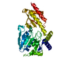
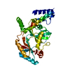
| Structure viewer | Molecule: MolmilJmol/JSmol |
|---|
- Downloads & links
-Download
| PDBx/mmCIF format | 1nml.cif.gz | 85.2 KB | Display | PDBx/mmCIF format |
|---|---|---|---|---|
| PDB format | pdb1nml.ent.gz | 64 KB | Display | PDB format |
| PDBx/mmJSON format | 1nml.json.gz | Tree view | PDBx/mmJSON format | |
| Others |  Other downloads Other downloads |
-Validation report
| Arichive directory | https://data.pdbj.org/pub/pdb/validation_reports/nm/1nmlftp://data.pdbj.org/pub/pdb/validation_reports/nm/1nml | HTTPS FTP |
|---|
-Related structure data
| Related structure data | 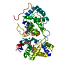 1rz5C 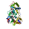 1rz6C 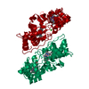 1eb7S S: Starting model for refinement C: citing same article ( |
|---|---|
| Similar structure data |


-Links
 PDBj
PDBj


- Assembly
| Deposited unit | 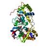
| ||||||||||
|---|---|---|---|---|---|---|---|---|---|---|---|
| 1 |
| ||||||||||
| 2 | 
| ||||||||||
| Unit cell |
|
-Components
| #1: Protein | Mass: 35383.512 Da / Num. of mol.: 1 / Source method: isolated from a natural source / Details: synonym Marinobacter hydrocarbonoclasticus Source: (natural) Marinobacter hydrocarbonoclasticus (bacteria)Strain: 617 / References: UniProt: P83787, cytochrome-c peroxidase | ||||||
|---|---|---|---|---|---|---|---|
| #2: Chemical |   Mass: 618.503 Da / Num. of mol.: 2 / Source method: obtained synthetically / Formula: C34H34FeN4O4 Mass: 618.503 Da / Num. of mol.: 2 / Source method: obtained synthetically / Formula: C34H34FeN4O4#3: Chemical | ChemComp-CIT /   Mass: 192.124 Da / Num. of mol.: 4 / Source method: obtained synthetically / Formula: C6H8O7 Mass: 192.124 Da / Num. of mol.: 4 / Source method: obtained synthetically / Formula: C6H8O7#4: Water | ChemComp-HOH / |  Mass: 18.015 Da / Num. of mol.: 319 / Source method: isolated from a natural source / Formula: H2O Mass: 18.015 Da / Num. of mol.: 319 / Source method: isolated from a natural source / Formula: H2OHas protein modification | Y | |
-Experimental details
-Experiment
| Experiment | Method: X-RAY DIFFRACTION / Number of used crystals: 1 |
|---|
- Sample preparation
| Crystal | Density Matthews: 4.26 Å3/Da / Density % sol: 70.9 % | ||||||||||||||||||||||||
|---|---|---|---|---|---|---|---|---|---|---|---|---|---|---|---|---|---|---|---|---|---|---|---|---|---|
| Crystal grow | Temperature: 277 K / Method: vapor diffusion / pH: 4 Details: sodium citrate, pH 4.0, VAPOR DIFFUSION, temperature 277K | ||||||||||||||||||||||||
| Crystal grow | *PLUS Temperature: 277 K / pH: 7.5 / Method: vapor diffusion, hanging drop / Details: Dias, J.M., (2002) Acta Cryst., D58, 697. | ||||||||||||||||||||||||
| Components of the solutions | *PLUS
|
-Data collection
| Diffraction | Mean temperature: 100 K |
|---|---|
| Diffraction source | Source: SYNCHROTRON / Site: ESRF  / Beamline: ID14-2 / Wavelength: 0.933 Å / Beamline: ID14-2 / Wavelength: 0.933 Å |
| Detector | Type: ADSC QUANTUM 4 / Detector: CCD / Date: Nov 18, 2000 |
| Radiation | Monochromator: 0.97 / Protocol: SINGLE WAVELENGTH / Monochromatic (M) / Laue (L): M / Scattering type: x-ray |
| Radiation wavelength | Wavelength: 0.933 Å / Relative weight: 1 |
| Reflection | Resolution: 2.2→20 Å / Num. all: 35161 / Num. obs: 35072 / % possible obs: 99.9 % / Observed criterion σ(I): -3 / Redundancy: 5 % / Biso Wilson estimate: 45.6 Å2 / Rsym value: 0.055 / Net I/σ(I): 26.5 |
| Reflection shell | Resolution: 2.2→2.28 Å / Redundancy: 5 % / Mean I/σ(I) obs: 4 / Num. unique all: 3475 / Rsym value: 0.535 / % possible all: 100 |
| Reflection | *PLUS Lowest resolution: 25 Å / Num. obs: 35359 / Num. measured all: 494949 / Rmerge(I) obs: 0.038 |
| Reflection shell | *PLUS % possible obs: 100 % / Rmerge(I) obs: 0.292 / Mean I/σ(I) obs: 7.2 |
- Processing
| Software |
| ||||||||||||||||||||||||||||
|---|---|---|---|---|---|---|---|---|---|---|---|---|---|---|---|---|---|---|---|---|---|---|---|---|---|---|---|---|---|
| Refinement | Method to determine structure: MOLECULAR REPLACEMENT Starting model: PDB ENTRY 1EB7 Resolution: 2.2→20 Å / Cross valid method: Rfree / σ(F): 0 / σ(I): 0 / Stereochemistry target values: Engh & Huber
| ||||||||||||||||||||||||||||
| Refinement step | Cycle: LAST / Resolution: 2.2→20 Å
| ||||||||||||||||||||||||||||
| Refine LS restraints |
| ||||||||||||||||||||||||||||
| Refinement | *PLUS Num. reflection obs: 33291 / % reflection Rfree: 5 % | ||||||||||||||||||||||||||||
| Solvent computation | *PLUS | ||||||||||||||||||||||||||||
| Displacement parameters | *PLUS | ||||||||||||||||||||||||||||
| Refine LS restraints | *PLUS Type: c_angle_deg / Dev ideal: 1.45 |
