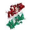
- PDB-1eb7: Crystal structure of the di-haem cytochrome c peroxidase from Pse... -
+
Open data
ID or keywords:
Loading...
-
Basic information
Entry
Database: PDB / ID: 1eb7
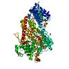
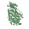
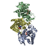
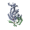
Title
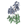
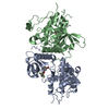
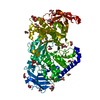
Crystal structure of the di-haem cytochrome c peroxidase from Pseudomonas aeruginosa
Components
CYTOCHROME C551 PEROXIDASE
Keywords
OXIDOREDUCTASE / PEROXIDASE / HEME / ELECTRON TRANSPORT
Function / homology
Function and homology information
cytochrome-c peroxidase / cytochrome-c peroxidase activity / electron transfer activity / periplasmic space / heme binding / metal ion binding Similarity search - Function
Di-c-type haem protein, MauG/cytochrome c peroxidase / Di-haem cytochrome c peroxidase / Di-haem cytochrome c peroxidase / : / Cytochrome c / Cytochrome c-like domain / Cytochrome Bc1 Complex; Chain D, domain 2 / Cytochrome c family profile. / Cytochrome c-like domain / Cytochrome c-like domain superfamily ...Di-c-type haem protein, MauG/cytochrome c peroxidase / Di-haem cytochrome c peroxidase / Di-haem cytochrome c peroxidase / : / Cytochrome c / Cytochrome c-like domain / Cytochrome Bc1 Complex; Chain D, domain 2 / Cytochrome c family profile. / Cytochrome c-like domain / Cytochrome c-like domain superfamily / Orthogonal Bundle / Mainly Alpha Similarity search - Domain/homology
Protocol: SINGLE WAVELENGTH / Monochromatic (M) / Laue (L): M / Scattering type: x-ray
Radiation wavelength
Wavelength: 1 Å / Relative weight: 1
Reflection
Resolution: 2.4→25 Å / Num. obs: 20541 / % possible obs: 96 % / Observed criterion σ(I): -3 / Redundancy: 3.9 % / Biso Wilson estimate: 40.6 Å2 / Rmerge(I) obs: 0.079 / Net I/σ(I): 18.5
Reflection
*PLUS
% possible obs: 96 % / Num. measured all: 80795
-
Processing
Software
Name
Version
Classification
X-PLOR
3.1
refinement
DENZO
datareduction
SCALEPACK
datascaling
CCP4
phasing
Refinement
Method to determine structure: MIR / Resolution: 2.4→25 Å / Isotropic thermal model: RESTRAINED / Cross valid method: THROUGHOUT / σ(F): 2 Details: RESIDUES 223-228 WERE DISORDERED AND NOT INCLUDED IN THE MODEL
In the structure databanks used in Yorodumi, some data are registered as the other names, "COVID-19 virus" and "2019-nCoV". Here are the details of the virus and the list of structure data.
Jan 31, 2019. EMDB accession codes are about to change! (news from PDBe EMDB page)
EMDB accession codes are about to change! (news from PDBe EMDB page)
The allocation of 4 digits for EMDB accession codes will soon come to an end. Whilst these codes will remain in use, new EMDB accession codes will include an additional digit and will expand incrementally as the available range of codes is exhausted. The current 4-digit format prefixed with “EMD-” (i.e. EMD-XXXX) will advance to a 5-digit format (i.e. EMD-XXXXX), and so on. It is currently estimated that the 4-digit codes will be depleted around Spring 2019, at which point the 5-digit format will come into force.
The EM Navigator/Yorodumi systems omit the EMD- prefix.
Related info.:Q: What is EMD? / ID/Accession-code notation in Yorodumi/EM Navigator
Yorodumi is a browser for structure data from EMDB, PDB, SASBDB, etc.
This page is also the successor to EM Navigator detail page, and also detail information page/front-end page for Omokage search.
The word "yorodu" (or yorozu) is an old Japanese word meaning "ten thousand". "mi" (miru) is to see.
Related info.:EMDB / PDB / SASBDB / Comparison of 3 databanks / Yorodumi Search / Aug 31, 2016. New EM Navigator & Yorodumi / Yorodumi Papers / Jmol/JSmol / Function and homology information / Changes in new EM Navigator and Yorodumi
 Movie
Movie Controller

 Yorodumi
 Open data
 Basic information
 Components
 Keywords
 Function and homology information
 PSEUDOMONAS AERUGINOSA (bacteria)
 X-RAY DIFFRACTION /
  Authors
 Citation
 Structure visualization
 Downloads & links
 Other downloads
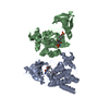


 PDBj
PDBj


 Assembly
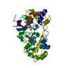

 Mass: 618.503 Da / Num. of mol.: 2 / Source method: obtained synthetically / Formula: C34H34FeN4O4
 Mass: 40.078 Da / Num. of mol.: 1 / Source method: obtained synthetically / Formula: Ca
 Mass: 18.015 Da / Num. of mol.: 73 / Source method: isolated from a natural source / Formula: H2O
 Sample preparation
 / Beamline: PX9.5 / Wavelength: 1
  Processing