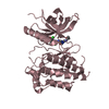
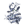
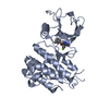
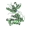
Entry Database : PDB / ID : 3gqlTitle Crystal Structure of activated receptor tyrosine kinase in complex with substrates Basic fibroblast growth factor receptor 1 Keywords / / / / / / / / / / / / / / / / / / / / / / Function / homology Function Domain/homology Component
/ / / / / / / / / / / / / / / / / / / / / / / / / / / / / / / / / / / / / / / / / / / / / / / / / / / / / / / / / / / / / / / / / / / / / / / / / / / / / / / / / / / / / / / / / / / / / / / / / / / / / / / / / / / / / / / / / / / / / / / / / / / / / Biological species Homo sapiens (human)Method / / / / Resolution : 2.8 Å Authors Bae, J.H. / Lew, E.D. / Yuzawa, S. / Tome, F. / Lax, I. / Schlessinger, J. Journal : Cell(Cambridge,Mass.) / Year : 2009Title : The selectivity of receptor tyrosine kinase signaling is controlled by a secondary SH2 domain binding site.Authors : Bae, J.H. / Lew, E.D. / Yuzawa, S. / Tome, F. / Lax, I. / Schlessinger, J. History Deposition Mar 24, 2009 Deposition site / Processing site Revision 1.0 Aug 18, 2009 Provider / Type Revision 1.1 Jul 13, 2011 Group Revision 1.2 Mar 21, 2012 Group Revision 1.3 Nov 1, 2017 Group / Category Revision 1.4 Oct 20, 2021 Group / Derived calculations / Category / struct_ref_seq_dif / struct_siteItem _database_2.pdbx_DOI / _database_2.pdbx_database_accession ... _database_2.pdbx_DOI / _database_2.pdbx_database_accession / _struct_ref_seq_dif.details / _struct_site.pdbx_auth_asym_id / _struct_site.pdbx_auth_comp_id / _struct_site.pdbx_auth_seq_id Revision 1.5 Feb 21, 2024 Group / Category / chem_comp_bond
Show all Show less
 Movie
Movie Controller

 Yorodumi
 Open data
 Basic information
 Components
 Keywords
 Function and homology information
 Homo sapiens (human)
 X-RAY DIFFRACTION /
  Authors
 Citation
 Structure visualization
 Downloads & links
 Other downloads
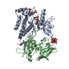

 PDBj
PDBj


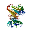
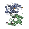
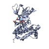
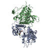
 Assembly
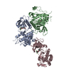

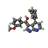
 Mass: 364.345 Da / Num. of mol.: 3 / Source method: obtained synthetically / Formula: C21H14F2N2O2
 Mass: 18.015 Da / Num. of mol.: 187 / Source method: isolated from a natural source / Formula: H2O
 Sample preparation
 / Beamline: X29A / Wavelength: 1.1
  Processing