+ Open data
- Basic information
| Entry | Database: PDB / ID: 6qk7 | |||||||||
|---|---|---|---|---|---|---|---|---|---|---|
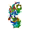
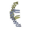
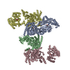
| Title | Elongator catalytic subcomplex Elp123 lobe | |||||||||
 Components Components |
| |||||||||
 Keywords Keywords | TRANSLATION / Elongator / yeast / tRNA modification / Elp123 | |||||||||
| Function / homology |  Function and homology information Function and homology informationtRNA uridine(34) acetyltransferase activity / tRNA carboxymethyluridine synthase / elongator holoenzyme complex / protein urmylation / tRNA wobble base 5-methoxycarbonylmethyl-2-thiouridinylation / tRNA wobble uridine modification / protein transport / regulation of translation / 4 iron, 4 sulfur cluster binding / microtubule binding ...tRNA uridine(34) acetyltransferase activity / tRNA carboxymethyluridine synthase / elongator holoenzyme complex / protein urmylation / tRNA wobble base 5-methoxycarbonylmethyl-2-thiouridinylation / tRNA wobble uridine modification / protein transport / regulation of translation / 4 iron, 4 sulfur cluster binding / microtubule binding / tRNA binding / regulation of transcription by RNA polymerase II / nucleoplasm / metal ion binding / identical protein binding / nucleus / cytosol / cytoplasm Similarity search - Function | |||||||||
| Biological species |  | |||||||||
| Method | ELECTRON MICROSCOPY / single particle reconstruction / cryo EM / Resolution: 3.3 Å | |||||||||
 Authors Authors | Dauden, M.I. / Jaciuk, M. / Glatt, S. | |||||||||
| Funding support |  Germany, Germany,  Poland, 2items Poland, 2items
| |||||||||
 Citation Citation |  Journal: Sci Adv / Year: 2019 Journal: Sci Adv / Year: 2019Title: Molecular basis of tRNA recognition by the Elongator complex. Authors: Maria I Dauden / Marcin Jaciuk / Felix Weis / Ting-Yu Lin / Carolin Kleindienst / Nour El Hana Abbassi / Heena Khatter / Rościsław Krutyhołowa / Karin D Breunig / Jan Kosinski / Christoph ...Authors: Maria I Dauden / Marcin Jaciuk / Felix Weis / Ting-Yu Lin / Carolin Kleindienst / Nour El Hana Abbassi / Heena Khatter / Rościsław Krutyhołowa / Karin D Breunig / Jan Kosinski / Christoph W Müller / Sebastian Glatt /   Abstract: The highly conserved Elongator complex modifies transfer RNAs (tRNAs) in their wobble base position, thereby regulating protein synthesis and ensuring proteome stability. The precise mechanisms of ...The highly conserved Elongator complex modifies transfer RNAs (tRNAs) in their wobble base position, thereby regulating protein synthesis and ensuring proteome stability. The precise mechanisms of tRNA recognition and its modification reaction remain elusive. Here, we show cryo-electron microscopy structures of the catalytic subcomplex of Elongator and its tRNA-bound state at resolutions of 3.3 and 4.4 Å. The structures resolve details of the catalytic site, including the substrate tRNA, the iron-sulfur cluster, and a SAM molecule, which are all validated by mutational analyses in vitro and in vivo. tRNA binding induces conformational rearrangements, which precisely position the targeted anticodon base in the active site. Our results provide the molecular basis for substrate recognition of Elongator, essential to understand its cellular function and role in neurodegenerative diseases and cancer. | |||||||||
| History |
|
- Structure visualization
| Movie |
 Movie viewer Movie viewer |
|---|---|
| Structure viewer | Molecule:  Molmil Molmil Jmol/JSmol Jmol/JSmol |
- Downloads & links
- Download
| PDBx/mmCIF format |  6qk7.cif.gz 6qk7.cif.gz | 491.3 KB | Display |  PDBx/mmCIF format PDBx/mmCIF format |
|---|---|---|---|---|
| PDB format |  pdb6qk7.ent.gz pdb6qk7.ent.gz | 380.4 KB | Display |  PDB format PDB format |
| PDBx/mmJSON format |  6qk7.json.gz 6qk7.json.gz | Tree view |  PDBx/mmJSON format PDBx/mmJSON format | |
| Others |  Other downloads Other downloads |
-Validation report
| Summary document |  6qk7_validation.pdf.gz 6qk7_validation.pdf.gz | 1.1 MB | Display |  wwPDB validaton report wwPDB validaton report |
|---|---|---|---|---|
| Full document |  6qk7_full_validation.pdf.gz 6qk7_full_validation.pdf.gz | 1.2 MB | Display | |
| Data in XML |  6qk7_validation.xml.gz 6qk7_validation.xml.gz | 79 KB | Display | |
| Data in CIF |  6qk7_validation.cif.gz 6qk7_validation.cif.gz | 119.1 KB | Display | |
| Arichive directory |  https://data.pdbj.org/pub/pdb/validation_reports/qk/6qk7 https://data.pdbj.org/pub/pdb/validation_reports/qk/6qk7 ftp://data.pdbj.org/pub/pdb/validation_reports/qk/6qk7 ftp://data.pdbj.org/pub/pdb/validation_reports/qk/6qk7 | HTTPS FTP |
-Related structure data
| Related structure data |  4571MC  4573C  4574C  4576C M: map data used to model this data C: citing same article ( |
|---|---|
| Similar structure data |
- Links
Links
- Assembly
| Deposited unit | 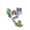
|
|---|---|
| 1 |
|
- Components
| #1: Protein | Mass: 153166.266 Da / Num. of mol.: 2 / Source method: isolated from a natural source Details: Chain D corresponds to the C-terminal domain of Elp1 Source: (natural)  Strain: ATCC 204508 / S288c / References: UniProt: Q06706 #2: Protein | | Mass: 89519.430 Da / Num. of mol.: 1 / Source method: isolated from a natural source Source: (natural)  Strain: ATCC 204508 / S288c / References: UniProt: P42935 #3: Protein | | Mass: 63755.059 Da / Num. of mol.: 1 / Source method: isolated from a natural source / Details: FeS cluster and 5DA Source: (natural)  Strain: ATCC 204508 / S288c / References: UniProt: Q02908, histone acetyltransferase #4: Chemical | ChemComp-SF4 / | #5: Chemical | ChemComp-5AD / | |
|---|
-Experimental details
-Experiment
| Experiment | Method: ELECTRON MICROSCOPY |
|---|---|
| EM experiment | Aggregation state: PARTICLE / 3D reconstruction method: single particle reconstruction |
- Sample preparation
| Component | Name: Elongator catalytic subcomplex Elp123 / Type: COMPLEX Details: The EM map corresponds to one lobe of the Elp123 complex, that includes one copy of Elp1, Elp2 and Elp3, and the C-terminal part of a second copy of Elp1. Entity ID: #1-#3 / Source: NATURAL | ||||||||||||||||||||||||||||||
|---|---|---|---|---|---|---|---|---|---|---|---|---|---|---|---|---|---|---|---|---|---|---|---|---|---|---|---|---|---|---|---|
| Molecular weight | Value: 0.621 MDa / Experimental value: NO | ||||||||||||||||||||||||||||||
| Source (natural) | Organism:  | ||||||||||||||||||||||||||||||
| Buffer solution | pH: 7.5 | ||||||||||||||||||||||||||||||
| Buffer component |
| ||||||||||||||||||||||||||||||
| Specimen | Conc.: 0.4 mg/ml / Embedding applied: NO / Shadowing applied: NO / Staining applied: NO / Vitrification applied: YES Details: The sample was cross-linked with 0.01% glutaraldehyde, quenched and then plunged. | ||||||||||||||||||||||||||||||
| Specimen support | Details: Pelco EasyGlow glow discharger, 20 mA / Grid material: COPPER / Grid mesh size: 200 divisions/in. / Grid type: Quantifoil R2/1 | ||||||||||||||||||||||||||||||
| Vitrification | Instrument: FEI VITROBOT MARK IV / Cryogen name: ETHANE / Humidity: 100 % / Chamber temperature: 277 K Details: 2.5 ul of sample, blotting parameters: wait time 15 s, blot force 5, blot time 5-8 s. |
- Electron microscopy imaging
| Experimental equipment |  Model: Titan Krios / Image courtesy: FEI Company |
|---|---|
| Microscopy | Model: FEI TITAN KRIOS Details: Gatan Quantum energy filter and a K2 Summit direct detector |
| Electron gun | Electron source:  FIELD EMISSION GUN / Accelerating voltage: 300 kV / Illumination mode: FLOOD BEAM FIELD EMISSION GUN / Accelerating voltage: 300 kV / Illumination mode: FLOOD BEAM |
| Electron lens | Mode: BRIGHT FIELD / Nominal magnification: 105000 X / Nominal defocus max: 2500 nm / Nominal defocus min: 1000 nm |
| Image recording | Electron dose: 43 e/Å2 / Detector mode: SUPER-RESOLUTION / Film or detector model: GATAN K2 SUMMIT (4k x 4k) / Num. of real images: 4614 |
| Image scans | Movie frames/image: 40 |
- Processing
| EM software |
| ||||||||||||||||||||||||||||||||||||||||||||||||||
|---|---|---|---|---|---|---|---|---|---|---|---|---|---|---|---|---|---|---|---|---|---|---|---|---|---|---|---|---|---|---|---|---|---|---|---|---|---|---|---|---|---|---|---|---|---|---|---|---|---|---|---|
| Image processing | Details: The detector was operated in super resolution mode. | ||||||||||||||||||||||||||||||||||||||||||||||||||
| CTF correction | Type: PHASE FLIPPING AND AMPLITUDE CORRECTION | ||||||||||||||||||||||||||||||||||||||||||||||||||
| Particle selection | Num. of particles selected: 1000000 Details: Initially 8563 particles were manually selected using EMAN2 boxer swarm tool, and used as 2D templates for the autopicking procedure in relion, that yielded 1 million particles. | ||||||||||||||||||||||||||||||||||||||||||||||||||
| Symmetry | Point symmetry: C1 (asymmetric) | ||||||||||||||||||||||||||||||||||||||||||||||||||
| 3D reconstruction | Resolution: 3.3 Å / Resolution method: FSC 0.143 CUT-OFF / Num. of particles: 84135 / Num. of class averages: 2 / Symmetry type: POINT | ||||||||||||||||||||||||||||||||||||||||||||||||||
| Atomic model building |
| ||||||||||||||||||||||||||||||||||||||||||||||||||
| Atomic model building | Source name: PDB / Type: experimental model
|
 Movie
Movie Controller



 PDBj
PDBj


