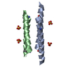
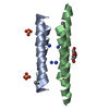
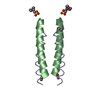
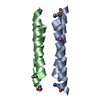
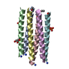
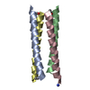
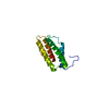
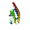
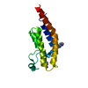
Entry Database : PDB / ID : 6q5qTitle Crystal structure of a CC-Hex mutant that forms an antiparallel four-helix coiled coil CC-Hex-KgEb CC-Hex-KgEb Keywords / / / / Function / homology Biological species synthetic construct (others) Method / / / Resolution : 1.08 Å Authors Zaccai, N.R. / Rhys, G.G. / Wood, C.W. / Beesley, J.L. / Brady, R.L. / Woolfson, D.N. Funding support Organization Grant number Country Biotechnology and Biological Sciences Research Council BB/J014400/1 Engineering and Physical Sciences Research Council EP/G036764/1 European Research Council 340764 Biotechnology and Biological Sciences Research Council BB/R00661X/1 Engineering and Physical Sciences Research Council EP/K03927X/1
Journal : J.Am.Chem.Soc. / Year : 2019Title : Navigating the Structural Landscape of De Novo alpha-Helical Bundles.Authors : Rhys, G.G. / Wood, C.W. / Beesley, J.L. / Zaccai, N.R. / Burton, A.J. / Brady, R.L. / Thomson, A.R. / Woolfson, D.N. History Deposition Dec 9, 2018 Deposition site / Processing site Revision 1.0 May 22, 2019 Provider / Type Revision 1.1 Jun 19, 2019 Group / Database references / Category / citation_author / pdbx_database_procItem _citation.journal_volume / _citation.page_first ... _citation.journal_volume / _citation.page_first / _citation.page_last / _citation.title / _citation_author.identifier_ORCID / _citation_author.name Revision 1.2 Nov 6, 2024 Group / Database references / Structure summaryCategory chem_comp_atom / chem_comp_bond ... chem_comp_atom / chem_comp_bond / database_2 / pdbx_entry_details / pdbx_modification_feature Item / _database_2.pdbx_database_accession
Show all Show less
 Movie
Movie Controller

 Yorodumi
 Open data
 Basic information
 Components
 Keywords
 Function and homology information
 X-RAY DIFFRACTION /
  Authors
 United Kingdom,
  Belgium, 5items
  Citation
 Structure visualization
 Downloads & links
 Other downloads
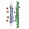


 PDBj
PDBj

 Assembly


 Mass: 150.087 Da / Num. of mol.: 1 / Source method: obtained synthetically / Formula: C4H6O6
 Mass: 18.015 Da / Num. of mol.: 34 / Source method: isolated from a natural source / Formula: H2O
 Sample preparation
 Processing