 Movie
Movie Controller

[English] 日本語
 Yorodumi
- PDB-6pw8: Hydrocarbon-Stapled Paxillin Peptide Bound to the Focal Adhesion ... -
+ Open data

- Basic information
| Entry | Database: PDB / ID: 6pw8 | |||||||||
|---|---|---|---|---|---|---|---|---|---|---|
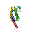
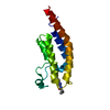
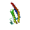
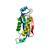
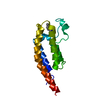
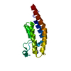
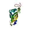
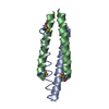
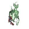
| Title | Hydrocarbon-Stapled Paxillin Peptide Bound to the Focal Adhesion Targeting (FAT) Domain of the Focal Adhesion Kinase (FAK) | |||||||||
 Components Components |
| |||||||||
 Keywords Keywords | PROTEIN BINDING/INHIBITOR / Inhibitor / Stapled Peptide / Tyrosine Kinase / PROTEIN BINDING-INHIBITOR complex | |||||||||
| Function / homology |  Function and homology information Function and homology informationnetrin-activated signaling pathway / regulation of substrate adhesion-dependent cell spreading / Regulation of cytoskeletal remodeling and cell spreading by IPP complex components / Localization of the PINCH-ILK-PARVIN complex to focal adhesions / regulation of endothelial cell migration / regulation of epithelial cell migration / Regulation of MITF-M-dependent genes involved in extracellular matrix, focal adhesion and epithelial-to-mesenchymal transition / detection of muscle stretch / positive regulation of cell-cell adhesion mediated by integrin / neuropilin binding ...netrin-activated signaling pathway / regulation of substrate adhesion-dependent cell spreading / Regulation of cytoskeletal remodeling and cell spreading by IPP complex components / Localization of the PINCH-ILK-PARVIN complex to focal adhesions / regulation of endothelial cell migration / regulation of epithelial cell migration / Regulation of MITF-M-dependent genes involved in extracellular matrix, focal adhesion and epithelial-to-mesenchymal transition / detection of muscle stretch / positive regulation of cell-cell adhesion mediated by integrin / neuropilin binding / vinculin binding / JUN kinase binding / signal complex assembly / negative regulation of cell adhesion mediated by integrin / positive regulation of macrophage proliferation / DCC mediated attractive signaling / Signal regulatory protein family interactions / positive regulation of fibroblast migration / microtubule associated complex / MET activates PTK2 signaling / growth hormone receptor signaling pathway / positive regulation of ubiquitin-dependent protein catabolic process / regulation of focal adhesion assembly / negative regulation of cell-cell adhesion / Apoptotic cleavage of cellular proteins / p130Cas linkage to MAPK signaling for integrins / positive regulation of wound healing / regulation of osteoblast differentiation / Fc-gamma receptor signaling pathway involved in phagocytosis / regulation of cytoskeleton organization / positive regulation of macrophage chemotaxis / establishment of cell polarity / vascular endothelial cell response to oscillatory fluid shear stress / GRB2:SOS provides linkage to MAPK signaling for Integrins / negative regulation of anoikis / positive regulation of epithelial cell migration / Estrogen-dependent nuclear events downstream of ESR-membrane signaling / ephrin receptor signaling pathway / vascular endothelial growth factor receptor signaling pathway / Smooth Muscle Contraction / GAB1 signalosome / RHO GTPases Activate WASPs and WAVEs / positive regulation of epithelial to mesenchymal transition / endothelial cell migration / regulation of cell adhesion / heart morphogenesis / PTK6 Regulates RHO GTPases, RAS GTPase and MAP kinases / positive regulation of stress fiber assembly / Integrin signaling / stress fiber / EPHB-mediated forward signaling / substrate adhesion-dependent cell spreading / protein tyrosine phosphatase activity / transforming growth factor beta receptor signaling pathway / NCAM signaling for neurite out-growth / SH2 domain binding / ciliary tip / molecular function activator activity / placenta development / Turbulent (oscillatory, disturbed) flow shear stress activates signaling by PIEZO1 and integrins in endothelial cells / axon guidance / integrin-mediated signaling pathway / FCGR3A-mediated phagocytosis / non-specific protein-tyrosine kinase / cellular response to reactive oxygen species / cell motility / non-membrane spanning protein tyrosine kinase activity / Regulation of actin dynamics for phagocytic cup formation / beta-catenin binding / VEGFA-VEGFR2 Pathway / integrin binding / epidermal growth factor receptor signaling pathway / cell-cell junction / regulation of cell shape / regulation of cell population proliferation / cell migration / lamellipodium / RAF/MAP kinase cascade / actin binding / protein tyrosine kinase activity / angiogenesis / protein phosphatase binding / cell cortex / cytoskeleton / positive regulation of phosphatidylinositol 3-kinase/protein kinase B signal transduction / cell adhesion / Extra-nuclear estrogen signaling / cilium / positive regulation of cell migration / ciliary basal body / focal adhesion / positive regulation of cell population proliferation / centrosome / protein kinase binding / negative regulation of apoptotic process / perinuclear region of cytoplasm / signal transduction / ATP binding / metal ion binding / nucleus Similarity search - Function | |||||||||
| Biological species |  Homo sapiens (human) Homo sapiens (human) | |||||||||
| Method |  X-RAY DIFFRACTION / SYNCHROTRON / MOLECULAR REPLACEMENT / Resolution: 1.95 Å X-RAY DIFFRACTION / SYNCHROTRON / MOLECULAR REPLACEMENT / Resolution: 1.95 Å | |||||||||
 Authors Authors | Thifault, D.G. / Fromme, P. / Martin-Garcia, J.M. | |||||||||
 Citation Citation | Journal: To Be Published Title: Stapled Peptide Ligand Bound to the Focal Adhesion Targeting (FAT) Domain of the Focal Adhesion Kinase (FAK) Authors: Thifault, D.G. / Fromme, P. / Martin-Garcia, J.M. #1: Journal: Acta Crystallogr.,Sect.D / Year: 2012 Title: Towards automated crystallographic structure refinement with phenix.refine. Authors: Afonine, P.V. / Grosse-Kunstleve, R.W. / Echols, N. / Headd, J.J. / Moriarty, N.W. / Mustyakimov, M. / Terwilliger, T.C. / Urzhumtsev, A. / Zwart, P.H. / Adams, P.D. #2: Journal: Acta Crystallogr D Biol Crystallogr / Year: 2010 Title: PHENIX: a comprehensive Python-based system for macromolecular structure solution. Authors: Paul D Adams / Pavel V Afonine / Gábor Bunkóczi / Vincent B Chen / Ian W Davis / Nathaniel Echols / Jeffrey J Headd / Li-Wei Hung / Gary J Kapral / Ralf W Grosse-Kunstleve / Airlie J McCoy ...Authors: Paul D Adams / Pavel V Afonine / Gábor Bunkóczi / Vincent B Chen / Ian W Davis / Nathaniel Echols / Jeffrey J Headd / Li-Wei Hung / Gary J Kapral / Ralf W Grosse-Kunstleve / Airlie J McCoy / Nigel W Moriarty / Robert Oeffner / Randy J Read / David C Richardson / Jane S Richardson / Thomas C Terwilliger / Peter H Zwart /  Abstract: Macromolecular X-ray crystallography is routinely applied to understand biological processes at a molecular level. However, significant time and effort are still required to solve and complete many ...Macromolecular X-ray crystallography is routinely applied to understand biological processes at a molecular level. However, significant time and effort are still required to solve and complete many of these structures because of the need for manual interpretation of complex numerical data using many software packages and the repeated use of interactive three-dimensional graphics. PHENIX has been developed to provide a comprehensive system for macromolecular crystallographic structure solution with an emphasis on the automation of all procedures. This has relied on the development of algorithms that minimize or eliminate subjective input, the development of algorithms that automate procedures that are traditionally performed by hand and, finally, the development of a framework that allows a tight integration between the algorithms. | |||||||||
| History |
|
- Structure visualization
| Structure viewer | Molecule: MolmilJmol/JSmol |
|---|
- Downloads & links
-Download
| PDBx/mmCIF format | 6pw8.cif.gz | 47 KB | Display | PDBx/mmCIF format |
|---|---|---|---|---|
| PDB format | pdb6pw8.ent.gz | 29.2 KB | Display | PDB format |
| PDBx/mmJSON format | 6pw8.json.gz | Tree view | PDBx/mmJSON format | |
| Others |  Other downloads Other downloads |
-Validation report
| Arichive directory | https://data.pdbj.org/pub/pdb/validation_reports/pw/6pw8ftp://data.pdbj.org/pub/pdb/validation_reports/pw/6pw8 | HTTPS FTP |
|---|
-Related structure data
| Related structure data |  1k05S S: Starting model for refinement |
|---|---|
| Similar structure data |

-Links
 PDBj
PDBj


- Assembly
| Deposited unit | 
| ||||||||||||
|---|---|---|---|---|---|---|---|---|---|---|---|---|---|
| 1 |
| ||||||||||||
| Unit cell |
|
-Components
| #1: Protein | Mass: 14018.440 Da / Num. of mol.: 1 Fragment: focal adhesion targeting domain (UNP residues 749-874) Source method: isolated from a genetically manipulated source Source: (gene. exp.) Homo sapiens (human) / Gene: PTK2, FAK, FAK1 / Production host:  References: UniProt: Q05397, non-specific protein-tyrosine kinase |
|---|---|
| #2: Protein/peptide | Mass: 1660.996 Da / Num. of mol.: 1 / Source method: obtained synthetically / Source: (synth.) Homo sapiens (human) / References: UniProt: P49023*PLUS |
| #3: Chemical | ChemComp-ZN /   Mass: 65.409 Da / Num. of mol.: 1 / Source method: obtained synthetically / Formula: Zn Mass: 65.409 Da / Num. of mol.: 1 / Source method: obtained synthetically / Formula: Zn |
| #4: Chemical | ChemComp-CL /   Mass: 35.453 Da / Num. of mol.: 1 / Source method: obtained synthetically / Formula: Cl Mass: 35.453 Da / Num. of mol.: 1 / Source method: obtained synthetically / Formula: Cl |
| #5: Water | ChemComp-HOH /  Mass: 18.015 Da / Num. of mol.: 92 / Source method: isolated from a natural source / Formula: H2O Mass: 18.015 Da / Num. of mol.: 92 / Source method: isolated from a natural source / Formula: H2O |
| Has ligand of interest | Y |
-Experimental details
-Experiment
| Experiment | Method: X-RAY DIFFRACTION / Number of used crystals: 1 |
|---|
- Sample preparation
| Crystal | Density Matthews: 2.15 Å3/Da / Density % sol: 42.85 % |
|---|---|
| Crystal grow | Temperature: 293 K / Method: vapor diffusion, hanging drop / Details: 0.02 M zinc chloride, 20% PEG3350 |
-Data collection
| Diffraction | Mean temperature: 293 K / Serial crystal experiment: N |
|---|---|
| Diffraction source | Source: SYNCHROTRON / Site: APS / Beamline: 23-ID-D / Wavelength: 1.02 Å |
| Detector | Type: DECTRIS PILATUS3 6M / Detector: PIXEL / Date: Apr 21, 2019 / Details: K-B Pair Bimorph Mirrors |
| Radiation | Monochromator: Si(111) / Protocol: SINGLE WAVELENGTH / Monochromatic (M) / Laue (L): M / Scattering type: x-ray |
| Radiation wavelength | Wavelength: 1.02 Å / Relative weight: 1 |
| Reflection | Resolution: 1.95→47.49 Å / Num. obs: 10290 / % possible obs: 99.7 % / Redundancy: 2 % / Biso Wilson estimate: 33.57 Å2 / Rmerge(I) obs: 0.114 / Net I/σ(I): 12.8 |
| Reflection shell | Resolution: 1.95→2.02 Å / Rmerge(I) obs: 1.859 / Num. unique obs: 1014 / % possible all: 99.5 |
- Processing
| Software |
| |||||||||||||||||||||||||||||||||||
|---|---|---|---|---|---|---|---|---|---|---|---|---|---|---|---|---|---|---|---|---|---|---|---|---|---|---|---|---|---|---|---|---|---|---|---|---|
| Refinement | Method to determine structure: MOLECULAR REPLACEMENT Starting model: PDB entry 1K05 Resolution: 1.95→37.66 Å / SU ML: 0.2365 / Cross valid method: THROUGHOUT / σ(F): 1.35 / Phase error: 29.0201 / Stereochemistry target values: CDL v1.2
| |||||||||||||||||||||||||||||||||||
| Solvent computation | Shrinkage radii: 0.9 Å / VDW probe radii: 1.11 Å / Solvent model: FLAT BULK SOLVENT MODEL | |||||||||||||||||||||||||||||||||||
| Displacement parameters | Biso mean: 38.06 Å2 | |||||||||||||||||||||||||||||||||||
| Refinement step | Cycle: LAST / Resolution: 1.95→37.66 Å
| |||||||||||||||||||||||||||||||||||
| Refine LS restraints |
| |||||||||||||||||||||||||||||||||||
| LS refinement shell |
|
