 Movie
Movie Controller

[English] 日本語
 Yorodumi
- PDB-6puv: Crystal Structure of the Carbohydrate Recognition Domain of the H... -
+ Open data

- Basic information
| Entry | Database: PDB / ID: 6puv | |||||||||||||||
|---|---|---|---|---|---|---|---|---|---|---|---|---|---|---|---|---|
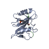
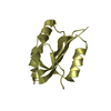
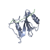
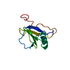
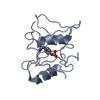
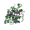
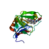
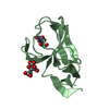
| Title | Crystal Structure of the Carbohydrate Recognition Domain of the Human Macrophage Galactose C-Type Lectin | |||||||||||||||
 Components Components | C-type lectin domain family 10 member A | |||||||||||||||
 Keywords Keywords | SIGNALING PROTEIN / C-TYPE LECTIN CRD | |||||||||||||||
| Function / homology |  Function and homology information Function and homology informationfucose binding / Dectin-2 family / pattern recognition receptor activity / D-mannose binding / endocytosis / carbohydrate binding / early endosome membrane / adaptive immune response / immune response / external side of plasma membrane ...fucose binding / Dectin-2 family / pattern recognition receptor activity / D-mannose binding / endocytosis / carbohydrate binding / early endosome membrane / adaptive immune response / immune response / external side of plasma membrane / innate immune response / lysosomal membrane / plasma membrane Similarity search - Function | |||||||||||||||
| Biological species |  Homo sapiens (human) Homo sapiens (human) | |||||||||||||||
| Method |  X-RAY DIFFRACTION / SYNCHROTRON / MOLECULAR REPLACEMENT / Resolution: 1.2 Å X-RAY DIFFRACTION / SYNCHROTRON / MOLECULAR REPLACEMENT / Resolution: 1.2 Å | |||||||||||||||
 Authors Authors | Birrane, G. / Murphy, P.V. / Gabba, A. / Luz, J.G. | |||||||||||||||
| Funding support |  Ireland, European Union, 4items Ireland, European Union, 4items
| |||||||||||||||
 Citation Citation | Journal: Biochemistry / Year: 2021 Title: Crystal Structure of the Carbohydrate Recognition Domain of the Human Macrophage Galactose C-Type Lectin Bound to GalNAc and the Tumor-Associated Tn Antigen. Authors: Gabba, A. / Bogucka, A. / Luz, J.G. / Diniz, A. / Coelho, H. / Corzana, F. / Canada, F.J. / Marcelo, F. / Murphy, P.V. / Birrane, G. | |||||||||||||||
| History |
|
- Structure visualization
| Structure viewer | Molecule: MolmilJmol/JSmol |
|---|
- Downloads & links
-Download
| PDBx/mmCIF format | 6puv.cif.gz | 70.6 KB | Display | PDBx/mmCIF format |
|---|---|---|---|---|
| PDB format | pdb6puv.ent.gz | 50.5 KB | Display | PDB format |
| PDBx/mmJSON format | 6puv.json.gz | Tree view | PDBx/mmJSON format | |
| Others |  Other downloads Other downloads |
-Validation report
| Arichive directory | https://data.pdbj.org/pub/pdb/validation_reports/pu/6puvftp://data.pdbj.org/pub/pdb/validation_reports/pu/6puv | HTTPS FTP |
|---|
-Related structure data
| Related structure data |  6py1C  6w12C  6xiyC  1dv8S S: Starting model for refinement C: citing same article ( |
|---|---|
| Similar structure data |


-Links
 PDBj
PDBj


- Assembly
| Deposited unit | 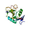
| ||||||||
|---|---|---|---|---|---|---|---|---|---|
| 1 |
| ||||||||
| Unit cell |
|
-Components
| #1: Protein | Mass: 14842.164 Da / Num. of mol.: 1 Source method: isolated from a genetically manipulated source Source: (gene. exp.) Homo sapiens (human) / Gene: CLEC10A, CLECSF13, CLECSF14, HML / Production host:  |
|---|---|
| #2: Chemical | ChemComp-CA /   Mass: 40.078 Da / Num. of mol.: 1 / Source method: obtained synthetically / Formula: Ca Mass: 40.078 Da / Num. of mol.: 1 / Source method: obtained synthetically / Formula: Ca |
| #3: Water | ChemComp-HOH /  Mass: 18.015 Da / Num. of mol.: 103 / Source method: isolated from a natural source / Formula: H2O Mass: 18.015 Da / Num. of mol.: 103 / Source method: isolated from a natural source / Formula: H2O |
| Has ligand of interest | N |
| Has protein modification | Y |
-Experimental details
-Experiment
| Experiment | Method: X-RAY DIFFRACTION / Number of used crystals: 1 |
|---|
- Sample preparation
| Crystal | Density Matthews: 1.96 Å3/Da / Density % sol: 37.29 % |
|---|---|
| Crystal grow | Temperature: 291 K / Method: vapor diffusion, sitting drop / pH: 5.5 / Details: 20% PEG 3000, 100mM Sodium citrate tribasic / PH range: 5.5 - 5.5 |
-Data collection
| Diffraction | Mean temperature: 125 K / Serial crystal experiment: N |
|---|---|
| Diffraction source | Source: SYNCHROTRON / Site: ESRF  / Beamline: ID30B / Wavelength: 0.9763 Å / Beamline: ID30B / Wavelength: 0.9763 Å |
| Detector | Type: DECTRIS PILATUS3 6M / Detector: PIXEL / Date: Jul 9, 2019 / Details: BE CRL/SI ELLIPTICAL MIRROR |
| Radiation | Monochromator: SI(111) / Protocol: SINGLE WAVELENGTH / Monochromatic (M) / Laue (L): M / Scattering type: x-ray |
| Radiation wavelength | Wavelength: 0.9763 Å / Relative weight: 1 |
| Reflection | Resolution: 1.2→45 Å / Num. obs: 34612 / % possible obs: 96.8 % / Redundancy: 3.7 % / CC1/2: 0.993 / Rpim(I) all: 0.037 / Rsym value: 0.066 / Net I/σ(I): 14 |
| Reflection shell | Resolution: 1.2→1.22 Å / Redundancy: 3.6 % / Mean I/σ(I) obs: 2.1 / Num. unique obs: 1714 / CC1/2: 0.824 / Rpim(I) all: 0.25 / Rsym value: 0.44 / % possible all: 99.2 |
- Processing
| Software |
| ||||||||||||||||||||
|---|---|---|---|---|---|---|---|---|---|---|---|---|---|---|---|---|---|---|---|---|---|
| Refinement | Method to determine structure: MOLECULAR REPLACEMENT Starting model: 1DV8 Resolution: 1.2→45 Å / Cross valid method: FREE R-VALUE
| ||||||||||||||||||||
| Refinement step | Cycle: LAST / Resolution: 1.2→45 Å
|
