 Movie
Movie Controller

[English] 日本語
 Yorodumi
- PDB-6py1: Crystal Structure of the Carbohydrate Recognition Domain of the H... -
+ Open data

- Basic information
| Entry | Database: PDB / ID: 6py1 | ||||||||||||
|---|---|---|---|---|---|---|---|---|---|---|---|---|---|
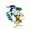
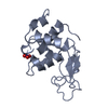
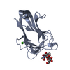
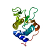
| Title | Crystal Structure of the Carbohydrate Recognition Domain of the Human Macrophage Galactose C-Type Lectin Bound to GalNAc | ||||||||||||
 Components Components | C-type lectin domain family 10 member A | ||||||||||||
 Keywords Keywords | SIGNALING PROTEIN / CRD | ||||||||||||
| Function / homology |  Function and homology information Function and homology informationfucose binding / Dectin-2 family / pattern recognition receptor activity / D-mannose binding / endocytosis / carbohydrate binding / early endosome membrane / adaptive immune response / immune response / external side of plasma membrane ...fucose binding / Dectin-2 family / pattern recognition receptor activity / D-mannose binding / endocytosis / carbohydrate binding / early endosome membrane / adaptive immune response / immune response / external side of plasma membrane / innate immune response / lysosomal membrane / plasma membrane Similarity search - Function | ||||||||||||
| Biological species |  Homo sapiens (human) Homo sapiens (human) | ||||||||||||
| Method |  X-RAY DIFFRACTION / SYNCHROTRON / MOLECULAR REPLACEMENT / Resolution: 1.701 Å X-RAY DIFFRACTION / SYNCHROTRON / MOLECULAR REPLACEMENT / Resolution: 1.701 Å | ||||||||||||
 Authors Authors | Birrane, G. / Murphy, P.V. / Gabba, A. / Luz, J.G. | ||||||||||||
| Funding support |  Ireland, European Union, 3items Ireland, European Union, 3items
| ||||||||||||
 Citation Citation | Journal: Biochemistry / Year: 2021 Title: Crystal Structure of the Carbohydrate Recognition Domain of the Human Macrophage Galactose C-Type Lectin Bound to GalNAc and the Tumor-Associated Tn Antigen. Authors: Gabba, A. / Bogucka, A. / Luz, J.G. / Diniz, A. / Coelho, H. / Corzana, F. / Canada, F.J. / Marcelo, F. / Murphy, P.V. / Birrane, G. | ||||||||||||
| History |
|
- Structure visualization
| Structure viewer | Molecule: MolmilJmol/JSmol |
|---|
- Downloads & links
-Download
| PDBx/mmCIF format | 6py1.cif.gz | 50.6 KB | Display | PDBx/mmCIF format |
|---|---|---|---|---|
| PDB format | pdb6py1.ent.gz | 32.4 KB | Display | PDB format |
| PDBx/mmJSON format | 6py1.json.gz | Tree view | PDBx/mmJSON format | |
| Others |  Other downloads Other downloads |
-Validation report
| Arichive directory | https://data.pdbj.org/pub/pdb/validation_reports/py/6py1ftp://data.pdbj.org/pub/pdb/validation_reports/py/6py1 | HTTPS FTP |
|---|
-Related structure data
| Related structure data |  6puvC  6w12C  6xiyC  1dv8S S: Starting model for refinement C: citing same article ( |
|---|---|
| Similar structure data |


-Links
 PDBj
PDBj


- Assembly
| Deposited unit | 
| |||||||||||||||
|---|---|---|---|---|---|---|---|---|---|---|---|---|---|---|---|---|
| 1 |
| |||||||||||||||
| 2 | 
| |||||||||||||||
| Unit cell |
| |||||||||||||||
| Components on special symmetry positions |
|
-Components
-Protein / Sugars , 2 types, 2 molecules A
| #1: Protein | Mass: 15057.370 Da / Num. of mol.: 1 Source method: isolated from a genetically manipulated source Source: (gene. exp.) Homo sapiens (human) / Gene: CLEC10A, CLECSF13, CLECSF14, HML / Production host:  |
|---|---|
| #2: Sugar | ChemComp-A2G /  Type: D-saccharide, alpha linking / Mass: 221.208 Da / Num. of mol.: 1 Type: D-saccharide, alpha linking / Mass: 221.208 Da / Num. of mol.: 1Source method: isolated from a genetically manipulated source Formula: C8H15NO6 / Feature type: SUBJECT OF INVESTIGATION |
-Non-polymers , 4 types, 155 molecules 


| #3: Chemical | ChemComp-ACT /  Mass: 59.044 Da / Num. of mol.: 1 Mass: 59.044 Da / Num. of mol.: 1Source method: isolated from a genetically manipulated source Formula: C2H3O2 | ||||
|---|---|---|---|---|---|
| #4: Chemical | ChemComp-CA /  Mass: 40.078 Da / Num. of mol.: 4 / Source method: obtained synthetically / Formula: Ca Mass: 40.078 Da / Num. of mol.: 4 / Source method: obtained synthetically / Formula: Ca#5: Chemical | ChemComp-CL /  Mass: 35.453 Da / Num. of mol.: 5 / Source method: obtained synthetically / Formula: Cl Mass: 35.453 Da / Num. of mol.: 5 / Source method: obtained synthetically / Formula: Cl#6: Water | ChemComp-HOH / | Mass: 18.015 Da / Num. of mol.: 145 / Source method: isolated from a natural source / Formula: H2O |
-Details
| Has ligand of interest | Y |
|---|---|
| Has protein modification | Y |
-Experimental details
-Experiment
| Experiment | Method: X-RAY DIFFRACTION / Number of used crystals: 1 |
|---|
- Sample preparation
| Crystal | Density Matthews: 2.92 Å3/Da / Density % sol: 57.91 % |
|---|---|
| Crystal grow | Temperature: 291 K / Method: vapor diffusion, sitting drop / pH: 7 Details: 100mM Tris pH 6.0 - 7.0, 200mM Magnesium chloride, 2.0 - 2.8M sodium chloride PH range: 6.0 - 7.0 |
-Data collection
| Diffraction | Mean temperature: 125 K / Serial crystal experiment: N |
|---|---|
| Diffraction source | Source: SYNCHROTRON / Site: ESRF  / Beamline: ID30B / Wavelength: 0.9763 Å / Beamline: ID30B / Wavelength: 0.9763 Å |
| Detector | Type: DECTRIS PILATUS3 6M / Detector: PIXEL / Date: Jul 19, 2018 / Details: BE CRL/SI ELLIPTICAL MIRROR |
| Radiation | Monochromator: SI(III) / Protocol: SINGLE WAVELENGTH / Monochromatic (M) / Laue (L): M / Scattering type: x-ray |
| Radiation wavelength | Wavelength: 0.9763 Å / Relative weight: 1 |
| Reflection | Resolution: 1.7→50 Å / Num. obs: 20014 / % possible obs: 100 % / Redundancy: 8.6 % / CC1/2: 0.977 / Rpim(I) all: 0.102 / Net I/σ(I): 12.4 |
| Reflection shell | Resolution: 1.7→1.73 Å / Redundancy: 6.5 % / Mean I/σ(I) obs: 1.6 / Num. unique obs: 1004 / CC1/2: 0.266 / Rpim(I) all: 0.867 |
- Processing
| Software |
| ||||||||||||||||||||||||||||||||||||||||||||||||||||||||||||||||||||||||||||||||||||||||||||||||||||||||||||||||||||||||||||||||||||||||||||||||||||||||||||||||
|---|---|---|---|---|---|---|---|---|---|---|---|---|---|---|---|---|---|---|---|---|---|---|---|---|---|---|---|---|---|---|---|---|---|---|---|---|---|---|---|---|---|---|---|---|---|---|---|---|---|---|---|---|---|---|---|---|---|---|---|---|---|---|---|---|---|---|---|---|---|---|---|---|---|---|---|---|---|---|---|---|---|---|---|---|---|---|---|---|---|---|---|---|---|---|---|---|---|---|---|---|---|---|---|---|---|---|---|---|---|---|---|---|---|---|---|---|---|---|---|---|---|---|---|---|---|---|---|---|---|---|---|---|---|---|---|---|---|---|---|---|---|---|---|---|---|---|---|---|---|---|---|---|---|---|---|---|---|---|---|---|---|
| Refinement | Method to determine structure: MOLECULAR REPLACEMENT Starting model: 1DV8 Resolution: 1.701→41.74 Å / Cor.coef. Fo:Fc: 0.964 / Cor.coef. Fo:Fc free: 0.952 / WRfactor Rfree: 0.185 / WRfactor Rwork: 0.162 / SU B: 1.898 / SU ML: 0.061 / Average fsc free: 0.9368 / Average fsc work: 0.945 / Cross valid method: FREE R-VALUE / ESU R: 0.087 / ESU R Free: 0.085 Details: Hydrogens have been added in their riding positions
| ||||||||||||||||||||||||||||||||||||||||||||||||||||||||||||||||||||||||||||||||||||||||||||||||||||||||||||||||||||||||||||||||||||||||||||||||||||||||||||||||
| Solvent computation | Ion probe radii: 0.8 Å / Shrinkage radii: 0.8 Å / VDW probe radii: 1.2 Å / Solvent model: BABINET MODEL PLUS MASK | ||||||||||||||||||||||||||||||||||||||||||||||||||||||||||||||||||||||||||||||||||||||||||||||||||||||||||||||||||||||||||||||||||||||||||||||||||||||||||||||||
| Displacement parameters | Biso mean: 18.426 Å2
| ||||||||||||||||||||||||||||||||||||||||||||||||||||||||||||||||||||||||||||||||||||||||||||||||||||||||||||||||||||||||||||||||||||||||||||||||||||||||||||||||
| Refinement step | Cycle: LAST / Resolution: 1.701→41.74 Å
| ||||||||||||||||||||||||||||||||||||||||||||||||||||||||||||||||||||||||||||||||||||||||||||||||||||||||||||||||||||||||||||||||||||||||||||||||||||||||||||||||
| Refine LS restraints |
|
