[English] 日本語
Yorodumi
- PDB-1iiz: Crystal Structure of the Induced Antibacterial Protein from Tasar... -

+
Open data


ID or keywords:

Loading...

-
Basic information

Entry
Database: PDB / ID: 1iiz
TitleCrystal Structure of the Induced Antibacterial Protein from Tasar Silkworm, Antheraea mylitta
ComponentsLYSOZYME
KeywordsHYDROLASE
Function / homology
Function and homology information


cytolysis / metabolic process / lysozyme / lysozyme activity / defense response to bacterium
Similarity search - Function
Lysozyme - #10 / Glycoside hydrolase, family 22, lysozyme / Glycoside hydrolase family 22 domain / Glycosyl hydrolases family 22 (GH22) domain signature. / Glycoside hydrolase, family 22 / C-type lysozyme/alpha-lactalbumin family / Glycosyl hydrolases family 22 (GH22) domain profile. / Alpha-lactalbumin / lysozyme C / Lysozyme / Lysozyme-like domain superfamily ...Lysozyme - #10 / Glycoside hydrolase, family 22, lysozyme / Glycoside hydrolase family 22 domain / Glycosyl hydrolases family 22 (GH22) domain signature. / Glycoside hydrolase, family 22 / C-type lysozyme/alpha-lactalbumin family / Glycosyl hydrolases family 22 (GH22) domain profile. / Alpha-lactalbumin / lysozyme C / Lysozyme / Lysozyme-like domain superfamily / Orthogonal Bundle / Mainly Alpha
Similarity search - Domain/homology
Biological speciesAntheraea mylitta (butterflies/moths)
MethodX-RAY DIFFRACTION / MOLECULAR REPLACEMENT / Resolution: 2.4 Å
AuthorsJain, D. / Nair, D.T. / Swaminathan, G.J. / Abraham, E.G. / Nagaraju, J. / Salunke, D.M.
CitationJournal: J.Biol.Chem. / Year: 2001
Title: Structure of the induced antibacterial protein from tasar silkworm, Antheraea mylitta. Implications to molecular evolution.
Authors: Jain, D. / Nair, D.T. / Swaminathan, G.J. / Abraham, E.G. / Nagaraju, J. / Salunke, D.M.
History
DepositionApr 24, 2001Deposition site: RCSB / Processing site: RCSB
Revision 1.0Dec 12, 2001Provider: repository / Type: Initial release
Revision 1.1Apr 27, 2008Group: Version format compliance
Revision 1.2Jul 13, 2011Group: Version format compliance
Revision 1.3Aug 16, 2023Group: Data collection / Database references / Refinement description
Category: chem_comp_atom / chem_comp_bond ...chem_comp_atom / chem_comp_bond / database_2 / pdbx_initial_refinement_model
Item: _database_2.pdbx_DOI / _database_2.pdbx_database_accession

-
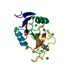
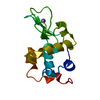
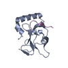
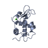
Structure visualization

Structure viewerMolecule:
MolmilJmol/JSmol

Downloads & links

-
Assembly

Deposited unit
A: LYSOZYME


Theoretical massNumber of molelcules
Total (without water)13,7611
Polymers13,7611
Non-polymers00
Water1,04558
1


  • Idetical with deposited unit
  • defined by author
TypeNameSymmetry operationNumber
identity operation1_555x,y,z1
Unit cell
Length a, b, c (Å)47.985, 51.700, 94.664
Angle α, β, γ (deg.)90.00, 90.00, 90.00
Int Tables number20
Space group name H-MC2221

-
Components

#1: Protein LYSOZYME /


Mass: 13761.416 Da / Num. of mol.: 1 / Source method: isolated from a natural source / Source: (natural) Antheraea mylitta (butterflies/moths) / References: UniProt: Q7SID7, lysozyme
#2: Water ChemComp-HOH / water / Water


Mass: 18.015 Da / Num. of mol.: 58 / Source method: isolated from a natural source / Formula: H2O

-
Experimental details

-
Experiment

ExperimentMethod: X-RAY DIFFRACTION

-
Sample preparation

CrystalDensity Matthews: 2.13 Å3/Da / Density % sol: 42.33 %
Crystal grow
*PLUS
Method: vapor diffusion, hanging drop
Components of the solutions
*PLUS
IDConc.Common nameCrystal-IDSol-IDDetails
10.1 Macetate1reservoirpH5.5
226.5 %PEG80001reservoir

-
Data collection

Diffraction sourceSource: ROTATING ANODE / Wavelength: 1.5418
DetectorType: MARRESEARCH / Detector: IMAGE PLATE
RadiationProtocol: SINGLE WAVELENGTH / Monochromatic (M) / Laue (L): M / Scattering type: x-ray
Radiation wavelengthWavelength: 1.5418 Å / Relative weight: 1
ReflectionBiso Wilson estimate: 16.8 Å2
Reflection
*PLUS
Highest resolution: 2.4 Å / Num. obs: 4807 / % possible obs: 98.7 % / Rmerge(I) obs: 0.083

-
Processing

Software
NameVersionClassification
DENZOdata reduction
SCALEPACKdata scaling
AMoREphasing
CNS0.5refinement
RefinementMethod to determine structure: MOLECULAR REPLACEMENT
Starting model: PDB ENTRY 1LMN
Resolution: 2.4→100 Å / Rfactor Rfree error: 0.013 / Data cutoff high absF: 227924.96 / Data cutoff low absF: 0 / Isotropic thermal model: RESTRAINED / Cross valid method: THROUGHOUT / σ(F): 0 / σ(I): 0
RfactorNum. reflection% reflectionSelection details
Rfree0.283 449 10.4 %RANDOM
Rwork0.231 ---
obs-4298 95.5 %-
Solvent computationSolvent model: FLAT MODEL / Bsol: 54.88 Å2 / ksol: 0.644 e/Å3
Displacement parametersBiso mean: 22.6 Å2
Baniso -1Baniso -2Baniso -3
1--0.36 Å20 Å20 Å2
2---3.65 Å20 Å2
3---4.02 Å2
Refine analyze
FreeObs
Luzzati coordinate error0.44 Å0.31 Å
Luzzati d res low-5 Å
Luzzati sigma a0.44 Å0.32 Å
Refinement stepCycle: LAST / Resolution: 2.4→100 Å
ProteinNucleic acidLigandSolventTotal
Num. atoms961 0 0 58 1019
Refine LS restraints
Refine-IDTypeDev idealDev ideal target
X-RAY DIFFRACTIONc_bond_d0.007
X-RAY DIFFRACTIONc_angle_deg1.6
X-RAY DIFFRACTIONc_dihedral_angle_d23.7
X-RAY DIFFRACTIONc_improper_angle_d0.77
X-RAY DIFFRACTIONc_mcbond_it5.261.5
X-RAY DIFFRACTIONc_mcangle_it8.162
X-RAY DIFFRACTIONc_scbond_it5.712
X-RAY DIFFRACTIONc_scangle_it8.352.5
LS refinement shellResolution: 2.4→2.54 Å / Rfactor Rfree error: 0.036 / Total num. of bins used: 6
RfactorNum. reflection% reflection
Rfree0.307 71 10.4 %
Rwork0.291 611 -
obs--92.2 %
Xplor file
Refine-IDSerial noParam fileTopol file
X-RAY DIFFRACTION1PROTEIN_REP.PARAMPROTEIN.TOP
X-RAY DIFFRACTION2WATER_REP.PARAMWATER_REP.TOP
Software
*PLUS
Name: CNS / Version: 0.5 / Classification: refinement
Refinement
*PLUS
Lowest resolution: 100 Å / σ(F): 0 / % reflection Rfree: 10.4 % / Rfactor obs: 0.238 / Rfactor Rfree: 0.27
Solvent computation
*PLUS
Displacement parameters
*PLUS
Biso mean: 22.6 Å2
Refine LS restraints
*PLUS
Refine-IDTypeDev idealDev ideal target
X-RAY DIFFRACTIONc_bond_d0.008
X-RAY DIFFRACTIONc_angle_deg1.7
X-RAY DIFFRACTIONc_dihedral_angle_d
X-RAY DIFFRACTIONc_dihedral_angle_deg23.7
X-RAY DIFFRACTIONc_improper_angle_d
X-RAY DIFFRACTIONc_improper_angle_deg0.77
X-RAY DIFFRACTIONc_mcbond_it5.261.5
X-RAY DIFFRACTIONc_scbond_it5.712
X-RAY DIFFRACTIONc_mcangle_it8.162
X-RAY DIFFRACTIONc_scangle_it8.352.5
LS refinement shell
*PLUS
Rfactor Rfree: 0.307 / % reflection Rfree: 10.4 % / Rfactor Rwork: 0.291

+
About Yorodumi

-
News

-
Feb 9, 2022. New format data for meta-information of EMDB entries

New format data for meta-information of EMDB entries

  • Version 3 of the EMDB header file is now the official format.
  • The previous official version 1.9 will be removed from the archive.

Related info.:EMDB header

External links:wwPDB to switch to version 3 of the EMDB data model

-
Aug 12, 2020. Covid-19 info

Covid-19 info

URL: https://pdbj.org/emnavi/covid19.php

New page: Covid-19 featured information page in EM Navigator.

Related info.:Covid-19 info / Mar 5, 2020. Novel coronavirus structure data

+
Mar 5, 2020. Novel coronavirus structure data

Novel coronavirus structure data

Related info.:Yorodumi Speices / Aug 12, 2020. Covid-19 info

External links:COVID-19 featured content - PDBj / Molecule of the Month (242):Coronavirus Proteases

+
Jan 31, 2019. EMDB accession codes are about to change! (news from PDBe EMDB page)

EMDB accession codes are about to change! (news from PDBe EMDB page)

  • The allocation of 4 digits for EMDB accession codes will soon come to an end. Whilst these codes will remain in use, new EMDB accession codes will include an additional digit and will expand incrementally as the available range of codes is exhausted. The current 4-digit format prefixed with “EMD-” (i.e. EMD-XXXX) will advance to a 5-digit format (i.e. EMD-XXXXX), and so on. It is currently estimated that the 4-digit codes will be depleted around Spring 2019, at which point the 5-digit format will come into force.
  • The EM Navigator/Yorodumi systems omit the EMD- prefix.

Related info.:Q: What is EMD? / ID/Accession-code notation in Yorodumi/EM Navigator

External links:EMDB Accession Codes are Changing Soon! / Contact to PDBj

+
Jul 12, 2017. Major update of PDB

Major update of PDB

  • wwPDB released updated PDB data conforming to the new PDBx/mmCIF dictionary.
  • This is a major update changing the version number from 4 to 5, and with Remediation, in which all the entries are updated.
  • In this update, many items about electron microscopy experimental information are reorganized (e.g. em_software).
  • Now, EM Navigator and Yorodumi are based on the updated data.

External links:wwPDB Remediation / Enriched Model Files Conforming to OneDep Data Standards Now Available in the PDB FTP Archive

-
Yorodumi

Thousand views of thousand structures

  • Yorodumi is a browser for structure data from EMDB, PDB, SASBDB, etc.
  • This page is also the successor to EM Navigator detail page, and also detail information page/front-end page for Omokage search.
  • The word "yorodu" (or yorozu) is an old Japanese word meaning "ten thousand". "mi" (miru) is to see.

Related info.:EMDB / PDB / SASBDB / Comparison of 3 databanks / Yorodumi Search / Aug 31, 2016. New EM Navigator & Yorodumi / Yorodumi Papers / Jmol/JSmol / Function and homology information / Changes in new EM Navigator and Yorodumi

Read more