 Movie
Movie Controller

[English] 日本語
 Yorodumi
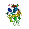
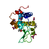
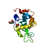
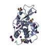
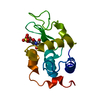
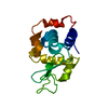
- PDB-1lmn: THE REFINED CRYSTAL STRUCTURE OF LYSOZYME FROM THE RAINBOW TROUT ... -
+ Open data

- Basic information
| Entry | Database: PDB / ID: 1lmn | |||||||||
|---|---|---|---|---|---|---|---|---|---|---|
| Title | THE REFINED CRYSTAL STRUCTURE OF LYSOZYME FROM THE RAINBOW TROUT (ONCORHYNCHUS MYKISS) | |||||||||
 Components Components | RAINBOW TROUT LYSOZYME | |||||||||
 Keywords Keywords | HYDROLASE (O-GLYCOSYL) | |||||||||
| Function / homology |  Function and homology information Function and homology informationlysozyme / lysozyme activity / killing of cells of another organism / defense response to Gram-negative bacterium / defense response to Gram-positive bacterium / : Similarity search - Function | |||||||||
| Biological species |  | |||||||||
| Method |  X-RAY DIFFRACTION / Resolution: 1.8 Å X-RAY DIFFRACTION / Resolution: 1.8 Å | |||||||||
 Authors Authors | Karlsen, S. / Hough, E. | |||||||||
 Citation Citation | Journal: Acta Crystallogr.,Sect.D / Year: 1995 Title: Refined crystal structure of lysozyme from the rainbow trout (Oncorhynchus mykiss). Authors: Karlsen, S. / Eliassen, B.E. / Hansen, L.K. / Larsen, R.L. / Riise, B.W. / Smalas, A.O. / Hough, E. / Grinde, B. #1: Journal: Eur.J.Biochem. / Year: 1988Title: Purification and Characterization of Two Lysozymes from Rainbow Trout (Salmo Gairdneri) Authors: Grinde, B. / Jolles, J. / Jolles, P. | |||||||||
| History |
|
- Structure visualization
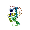
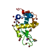
| Structure viewer | Molecule: MolmilJmol/JSmol |
|---|
- Downloads & links
-Download
| PDBx/mmCIF format | 1lmn.cif.gz | 40 KB | Display | PDBx/mmCIF format |
|---|---|---|---|---|
| PDB format | pdb1lmn.ent.gz | 27.5 KB | Display | PDB format |
| PDBx/mmJSON format | 1lmn.json.gz | Tree view | PDBx/mmJSON format | |
| Others |  Other downloads Other downloads |
-Validation report
| Arichive directory | https://data.pdbj.org/pub/pdb/validation_reports/lm/1lmnftp://data.pdbj.org/pub/pdb/validation_reports/lm/1lmn | HTTPS FTP |
|---|
-Related structure data
| Similar structure data |
|---|

-Links
 PDBj
PDBj- Assembly
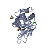
| Deposited unit | 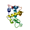
| ||||||||
|---|---|---|---|---|---|---|---|---|---|
| 1 |
| ||||||||
| Unit cell |
|
-Components
| #1: Protein | Mass: 14303.068 Da / Num. of mol.: 1 Source method: isolated from a genetically manipulated source Source: (gene. exp.) |
|---|---|
| #2: Water | ChemComp-HOH /  Mass: 18.015 Da / Num. of mol.: 127 / Source method: isolated from a natural source / Formula: H2O Mass: 18.015 Da / Num. of mol.: 127 / Source method: isolated from a natural source / Formula: H2O |
| Has protein modification | Y |
-Experimental details
-Experiment
| Experiment | Method: X-RAY DIFFRACTION |
|---|
- Sample preparation
| Crystal | Density Matthews: 3.23 Å3/Da / Density % sol: 61.93 % | ||||||||||||||||||||||||||||||||||||
|---|---|---|---|---|---|---|---|---|---|---|---|---|---|---|---|---|---|---|---|---|---|---|---|---|---|---|---|---|---|---|---|---|---|---|---|---|---|
| Crystal grow | *PLUS Temperature: 277 K / Method: vapor diffusion, hanging drop / PH range low: 10 / PH range high: 4 | ||||||||||||||||||||||||||||||||||||
| Components of the solutions | *PLUS
|
-Data collection
| Radiation | Scattering type: x-ray |
|---|---|
| Radiation wavelength | Relative weight: 1 |
| Reflection | Num. obs: 16305 / % possible obs: 91.2 % / Observed criterion σ(I): 3 |
| Reflection | *PLUS Highest resolution: 1.8 Å / Lowest resolution: 42.11 Å / Num. measured all: 70573 / Rmerge(I) obs: 0.0417 |
- Processing
| Software |
| ||||||||||||||||||||||||||||||||||||||||||||||||||||||||||||||||||||||||||||||||||||
|---|---|---|---|---|---|---|---|---|---|---|---|---|---|---|---|---|---|---|---|---|---|---|---|---|---|---|---|---|---|---|---|---|---|---|---|---|---|---|---|---|---|---|---|---|---|---|---|---|---|---|---|---|---|---|---|---|---|---|---|---|---|---|---|---|---|---|---|---|---|---|---|---|---|---|---|---|---|---|---|---|---|---|---|---|---|
| Refinement | Resolution: 1.8→8 Å / σ(F): 3 Details: THE STRUCTURE WAS SOLVED BY MOLECULAR REPLACEMENT METHODS USING THE MERLOT PACKAGE (FITZGERALD, P. (1988) J. APPL. CRYST. 21, 273-278) AND THE REFINED MODEL OF THE HEN EGG-WHITE LYSOZYME AS ...Details: THE STRUCTURE WAS SOLVED BY MOLECULAR REPLACEMENT METHODS USING THE MERLOT PACKAGE (FITZGERALD, P. (1988) J. APPL. CRYST. 21, 273-278) AND THE REFINED MODEL OF THE HEN EGG-WHITE LYSOZYME AS SEARCH MODEL (BROOKHAVEN PROTEIN DATA BANK, ENTRY 1LZT, HODSON ET AL.,(1990), ACTA. CRYST. B46, 52-62.
| ||||||||||||||||||||||||||||||||||||||||||||||||||||||||||||||||||||||||||||||||||||
| Displacement parameters | Biso mean: 24.4 Å2 | ||||||||||||||||||||||||||||||||||||||||||||||||||||||||||||||||||||||||||||||||||||
| Refinement step | Cycle: LAST / Resolution: 1.8→8 Å
| ||||||||||||||||||||||||||||||||||||||||||||||||||||||||||||||||||||||||||||||||||||
| Refine LS restraints |
| ||||||||||||||||||||||||||||||||||||||||||||||||||||||||||||||||||||||||||||||||||||
| Software | *PLUS Name: PROLSQ / Classification: refinement | ||||||||||||||||||||||||||||||||||||||||||||||||||||||||||||||||||||||||||||||||||||
| Refinement | *PLUS Rfactor obs: 0.174 | ||||||||||||||||||||||||||||||||||||||||||||||||||||||||||||||||||||||||||||||||||||
| Solvent computation | *PLUS | ||||||||||||||||||||||||||||||||||||||||||||||||||||||||||||||||||||||||||||||||||||
| Displacement parameters | *PLUS |
