 Movie
Movie Controller

[English] 日本語
 Yorodumi
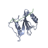
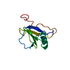
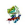
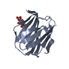
- PDB-1d4w: CRYSTAL STRUCTURE OF THE XLP PROTEIN SAP IN COMPLEX WITH SLAM PHO... -
+ Open data

- Basic information
| Entry | Database: PDB / ID: 1d4w | ||||||
|---|---|---|---|---|---|---|---|
| Title | CRYSTAL STRUCTURE OF THE XLP PROTEIN SAP IN COMPLEX WITH SLAM PHOSPHOPEPTIDE | ||||||
 Components Components |
| ||||||
 Keywords Keywords | SIGNALING PROTEIN / SH2 DOMAIN / PHOSPHOTYROSINE RECOGNIITON / PEPTIDE RECOGNITION / SIGNAL TRANSDUCTION / LYMPHOPROLIFERATIVE DISEASE | ||||||
| Function / homology |  Function and homology information Function and homology informationnatural killer cell proliferation / negative regulation of CD40 signaling pathway / negative regulation of T cell cytokine production / regulation of vesicle fusion / myeloid dendritic cell activation involved in immune response / leukocyte chemotaxis involved in inflammatory response / positive regulation of dendritic cell chemotaxis / positive regulation of T-helper 1 cell cytokine production / natural killer cell differentiation / negative regulation of interleukin-12 production ...natural killer cell proliferation / negative regulation of CD40 signaling pathway / negative regulation of T cell cytokine production / regulation of vesicle fusion / myeloid dendritic cell activation involved in immune response / leukocyte chemotaxis involved in inflammatory response / positive regulation of dendritic cell chemotaxis / positive regulation of T-helper 1 cell cytokine production / natural killer cell differentiation / negative regulation of interleukin-12 production / positive regulation of natural killer cell mediated cytotoxicity / natural killer cell activation / positive regulation of activated T cell proliferation / natural killer cell mediated cytotoxicity / positive regulation of macrophage chemotaxis / negative regulation of type II interferon production / humoral immune response / negative regulation of interleukin-6 production / negative regulation of tumor necrosis factor production / regulation of immune response / negative regulation of T cell receptor signaling pathway / antigen binding / cellular defense response / phagocytosis / phagocytic vesicle / SH2 domain binding / positive regulation of JNK cascade / Immunoregulatory interactions between a Lymphoid and a non-Lymphoid cell / positive regulation of type II interferon production / transmembrane signaling receptor activity / cell-cell signaling / virus receptor activity / signaling receptor activity / adaptive immune response / positive regulation of ERK1 and ERK2 cascade / protein-macromolecule adaptor activity / cell adhesion / immune response / external side of plasma membrane / innate immune response / positive regulation of cell population proliferation / extracellular exosome / identical protein binding / plasma membrane / cytosol / cytoplasm Similarity search - Function | ||||||
| Biological species |  Homo sapiens (human) Homo sapiens (human) | ||||||
| Method |  X-RAY DIFFRACTION / Resolution: 1.8 Å X-RAY DIFFRACTION / Resolution: 1.8 Å | ||||||
 Authors Authors | Poy, F. / Yaffe, M.B. / Sayos, J. / Saxena, K. / Eck, M.J. | ||||||
 Citation Citation | Journal: Mol.Cell / Year: 1999 Title: Crystal structures of the XLP protein SAP reveal a class of SH2 domains with extended, phosphotyrosine-independent sequence recognition. Authors: Poy, F. / Yaffe, M.B. / Sayos, J. / Saxena, K. / Morra, M. / Sumegi, J. / Cantley, L.C. / Terhorst, C. / Eck, M.J. #1: Journal: Nature / Year: 1998Title: The X-linked lymphoproliferative disease gene product SAP regulates signals induced through the co-receptor SLAM Authors: Sayos, J. / Wu, C. / Morra, M. / Wang, N. / Terhorst, C. | ||||||
| History |
|
- Structure visualization
| Structure viewer | Molecule: MolmilJmol/JSmol |
|---|
- Downloads & links
-Download
| PDBx/mmCIF format | 1d4w.cif.gz | 66.5 KB | Display | PDBx/mmCIF format |
|---|---|---|---|---|
| PDB format | pdb1d4w.ent.gz | 48.8 KB | Display | PDB format |
| PDBx/mmJSON format | 1d4w.json.gz | Tree view | PDBx/mmJSON format | |
| Others |  Other downloads Other downloads |
-Validation report
| Arichive directory | https://data.pdbj.org/pub/pdb/validation_reports/d4/1d4wftp://data.pdbj.org/pub/pdb/validation_reports/d4/1d4w | HTTPS FTP |
|---|
-Related structure data


-Links
 PDBj
PDBj
- Assembly
| Deposited unit | 
| ||||||||
|---|---|---|---|---|---|---|---|---|---|
| 1 | 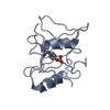
| ||||||||
| 2 | 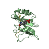
| ||||||||
| Unit cell |
|
-Components
| #1: Protein | Mass: 11702.393 Da / Num. of mol.: 2 / Fragment: SH2 DOMAIN / Mutation: RESIDUES 1-104 Source method: isolated from a genetically manipulated source Source: (gene. exp.) Homo sapiens (human) / Tissue: BLOOD / Cell: T CELL / Plasmid: T7-PRSET / Production host:  #2: Protein/peptide | Mass: 1360.491 Da / Num. of mol.: 2 / Fragment: CYTOPLASMIC TAIL SYNTHETIC PHOSPOPEPTIDE / Source method: obtained synthetically / Details: THIS PEPTIDE WAS CHEMICALLY SYNTHESIZED. / References: UniProt: Q13291 #3: Water | ChemComp-HOH / |  Mass: 18.015 Da / Num. of mol.: 413 / Source method: isolated from a natural source / Formula: H2O Mass: 18.015 Da / Num. of mol.: 413 / Source method: isolated from a natural source / Formula: H2OHas protein modification | Y | |
|---|
-Experimental details
-Experiment
| Experiment | Method: X-RAY DIFFRACTION / Number of used crystals: 1 |
|---|
- Sample preparation
| Crystal | Density Matthews: 2.6 Å3/Da / Density % sol: 52.77 % | ||||||||||||||||||||||||||||||||||||||||||||||||||||||
|---|---|---|---|---|---|---|---|---|---|---|---|---|---|---|---|---|---|---|---|---|---|---|---|---|---|---|---|---|---|---|---|---|---|---|---|---|---|---|---|---|---|---|---|---|---|---|---|---|---|---|---|---|---|---|---|
| Crystal grow | Temperature: 295 K / Method: vapor diffusion, hanging drop / pH: 5.6 Details: 30% PEG 8000, 20% GLYCEROL, 100 MM SODIUM CITRATE, 10MM DTT, pH 5.6, VAPOR DIFFUSION, HANGING DROP, temperature 22K | ||||||||||||||||||||||||||||||||||||||||||||||||||||||
| Crystal grow | *PLUS pH: 7.5 | ||||||||||||||||||||||||||||||||||||||||||||||||||||||
| Components of the solutions | *PLUS
|
-Data collection
| Diffraction | Mean temperature: 100 K |
|---|---|
| Diffraction source | Source: ROTATING ANODE / Type: RIGAKU RU300 / Wavelength: 1.5418 |
| Detector | Type: MARRESEARCH / Detector: IMAGE PLATE |
| Radiation | Protocol: SINGLE WAVELENGTH / Monochromatic (M) / Laue (L): M / Scattering type: x-ray |
| Radiation wavelength | Wavelength: 1.5418 Å / Relative weight: 1 |
| Reflection | Resolution: 1.8→20 Å / Num. all: 23946 / % possible obs: 94.8 % / Rmerge(I) obs: 0.076 |
| Reflection | *PLUS Num. obs: 23946 / Num. measured all: 113987 |
- Processing
| Software |
| |||||||||||||||||||||||||||||||||
|---|---|---|---|---|---|---|---|---|---|---|---|---|---|---|---|---|---|---|---|---|---|---|---|---|---|---|---|---|---|---|---|---|---|---|
| Refinement | Resolution: 1.8→10 Å / σ(F): 4 / Stereochemistry target values: ENGH& HUBER
| |||||||||||||||||||||||||||||||||
| Refinement step | Cycle: LAST / Resolution: 1.8→10 Å
| |||||||||||||||||||||||||||||||||
| Refine LS restraints |
| |||||||||||||||||||||||||||||||||
| Software | *PLUS Name: SHELXL-97 / Classification: refinement | |||||||||||||||||||||||||||||||||
| Refinement | *PLUS Highest resolution: 1.8 Å / Lowest resolution: 10 Å / σ(F): 4 / Rfactor Rfree: 0.24 | |||||||||||||||||||||||||||||||||
| Solvent computation | *PLUS | |||||||||||||||||||||||||||||||||
| Displacement parameters | *PLUS | |||||||||||||||||||||||||||||||||
| Refine LS restraints | *PLUS
|
