 Movie
Movie Controller

[English] 日本語
 Yorodumi
- PDB-5xor: Crystal structure of N-terminal replicase protein of porcine circ... -
+ Open data

- Basic information
| Entry | Database: PDB / ID: 5xor | ||||||
|---|---|---|---|---|---|---|---|
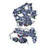
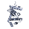
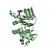
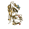
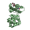
| Title | Crystal structure of N-terminal replicase protein of porcine circovirus type 2 | ||||||
 Components Components | Rep protein | ||||||
 Keywords Keywords | REPLICATION / PCV2 / Rep / Dimer | ||||||
| Function / homology |  Function and homology information Function and homology informationDNA endonuclease activity, producing 5'-phosphomonoesters / nucleotidyltransferase activity / DNA replication / RNA helicase activity / nucleotide binding / host cell nucleus / DNA binding / RNA binding / metal ion binding Similarity search - Function | ||||||
| Biological species |   Porcine circovirus 2 Porcine circovirus 2 | ||||||
| Method |  X-RAY DIFFRACTION / SYNCHROTRON / MOLECULAR REPLACEMENT / Resolution: 2.699 Å X-RAY DIFFRACTION / SYNCHROTRON / MOLECULAR REPLACEMENT / Resolution: 2.699 Å | ||||||
 Authors Authors | Song, Y. / Peng, G. | ||||||
 Citation Citation | Journal: J. Virol. / Year: 2018 Title: Crystal Structure of the Dimerized N Terminus of Porcine Circovirus Type 2 Replicase Protein Reveals a Novel Antiviral Interface Authors: Luo, G. / Zhu, X. / Lv, Y. / Lv, B. / Fang, J. / Cao, S. / Chen, H. / Peng, G. / Song, Y. | ||||||
| History |
|
- Structure visualization
| Structure viewer | Molecule: MolmilJmol/JSmol |
|---|
- Downloads & links
-Download
| PDBx/mmCIF format | 5xor.cif.gz | 115.6 KB | Display | PDBx/mmCIF format |
|---|---|---|---|---|
| PDB format | pdb5xor.ent.gz | 89.2 KB | Display | PDB format |
| PDBx/mmJSON format | 5xor.json.gz | Tree view | PDBx/mmJSON format | |
| Others |  Other downloads Other downloads |
-Validation report
| Arichive directory | https://data.pdbj.org/pub/pdb/validation_reports/xo/5xorftp://data.pdbj.org/pub/pdb/validation_reports/xo/5xor | HTTPS FTP |
|---|
-Related structure data
| Related structure data |  2hw0S S: Starting model for refinement |
|---|---|
| Similar structure data |


-Links
 PDBj
PDBj
- Assembly
| Deposited unit | 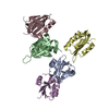
| ||||||||
|---|---|---|---|---|---|---|---|---|---|
| 1 | 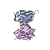
| ||||||||
| 2 | 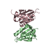
| ||||||||
| 3 | 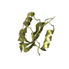
| ||||||||
| Unit cell |
|
-Components
| #1: Protein | Mass: 18077.359 Da / Num. of mol.: 5 / Fragment: UNP residues 1-150 Source method: isolated from a genetically manipulated source Source: (gene. exp.) Porcine circovirus 2Production host:  References: UniProt: A9YPG7 #2: Water | ChemComp-HOH / |  Mass: 18.015 Da / Num. of mol.: 64 / Source method: isolated from a natural source / Formula: H2O Mass: 18.015 Da / Num. of mol.: 64 / Source method: isolated from a natural source / Formula: H2O |
|---|
-Experimental details
-Experiment
| Experiment | Method: X-RAY DIFFRACTION / Number of used crystals: 1 |
|---|
- Sample preparation
| Crystal | Density Matthews: 2.13 Å3/Da / Density % sol: 42.38 % Description: THE ENTRY CONTAINS FRIEDEL PAIRS IN F_PLUS/MINUS COLUMNS. |
|---|---|
| Crystal grow | Temperature: 293 K / Method: evaporation Details: 0.1M HEPES sodium pH 8.0, 0.45 M sodium citrate tribasic dihydrate, 50% (+/-)-2-Methyl-2,4-pentanediol |
-Data collection
| Diffraction | Mean temperature: 100 K |
|---|---|
| Diffraction source | Source: SYNCHROTRON / Site: BSRF  / Beamline: 3W1A / Wavelength: 1 Å / Beamline: 3W1A / Wavelength: 1 Å |
| Detector | Type: MARMOSAIC 225 mm CCD / Detector: CCD / Date: Oct 28, 2013 |
| Radiation | Protocol: SINGLE WAVELENGTH / Monochromatic (M) / Laue (L): M / Scattering type: x-ray |
| Radiation wavelength | Wavelength: 1 Å / Relative weight: 1 |
| Reflection | Resolution: 2→50 Å / Num. obs: 46536 / % possible obs: 97.9 % / Redundancy: 4.4 % / Net I/σ(I): 12.2 |
- Processing
| Software |
| ||||||||||||||||||||||||||||||||||||||||||||||||||||||||||||||||||||||||||||||||||||||||||||||||||||||||||||||||||||||||||||||||||||||||||||||||||||||||||||||||||||||||||||||||||||||||||||||||||||
|---|---|---|---|---|---|---|---|---|---|---|---|---|---|---|---|---|---|---|---|---|---|---|---|---|---|---|---|---|---|---|---|---|---|---|---|---|---|---|---|---|---|---|---|---|---|---|---|---|---|---|---|---|---|---|---|---|---|---|---|---|---|---|---|---|---|---|---|---|---|---|---|---|---|---|---|---|---|---|---|---|---|---|---|---|---|---|---|---|---|---|---|---|---|---|---|---|---|---|---|---|---|---|---|---|---|---|---|---|---|---|---|---|---|---|---|---|---|---|---|---|---|---|---|---|---|---|---|---|---|---|---|---|---|---|---|---|---|---|---|---|---|---|---|---|---|---|---|---|---|---|---|---|---|---|---|---|---|---|---|---|---|---|---|---|---|---|---|---|---|---|---|---|---|---|---|---|---|---|---|---|---|---|---|---|---|---|---|---|---|---|---|---|---|---|---|---|---|
| Refinement | Method to determine structure: MOLECULAR REPLACEMENT Starting model: 2hw0 Resolution: 2.699→42.34 Å / SU ML: 0.43 / Cross valid method: THROUGHOUT / σ(F): 1.35 / Phase error: 32.62 / Stereochemistry target values: ML Details: SF FILE CONTAINS FRIEDEL PAIRS UNDER I/F_MINUS AND I/F_PLUS COLUMNS.
| ||||||||||||||||||||||||||||||||||||||||||||||||||||||||||||||||||||||||||||||||||||||||||||||||||||||||||||||||||||||||||||||||||||||||||||||||||||||||||||||||||||||||||||||||||||||||||||||||||||
| Solvent computation | Shrinkage radii: 0.9 Å / VDW probe radii: 1.11 Å / Solvent model: FLAT BULK SOLVENT MODEL | ||||||||||||||||||||||||||||||||||||||||||||||||||||||||||||||||||||||||||||||||||||||||||||||||||||||||||||||||||||||||||||||||||||||||||||||||||||||||||||||||||||||||||||||||||||||||||||||||||||
| Refinement step | Cycle: LAST / Resolution: 2.699→42.34 Å
| ||||||||||||||||||||||||||||||||||||||||||||||||||||||||||||||||||||||||||||||||||||||||||||||||||||||||||||||||||||||||||||||||||||||||||||||||||||||||||||||||||||||||||||||||||||||||||||||||||||
| Refine LS restraints |
| ||||||||||||||||||||||||||||||||||||||||||||||||||||||||||||||||||||||||||||||||||||||||||||||||||||||||||||||||||||||||||||||||||||||||||||||||||||||||||||||||||||||||||||||||||||||||||||||||||||
| LS refinement shell |
|
