National Institutes of Health/National Institute of General Medical Sciences (NIH/NIGMS)
GM105826
United States
National Institutes of Health/National Institute Of Allergy and Infectious Diseases (NIH/NIAID)
AI117675
United States
National Institutes of Health/National Institute Of Allergy and Infectious Diseases (NIH/NIAID)
AI084817
United States
Citation
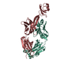
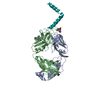
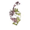
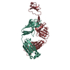
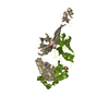
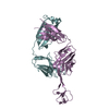
Journal: Sci Adv / Year: 2020 Title: Structural basis of broad HIV neutralization by a vaccine-induced cow antibody. Authors: Robyn L Stanfield / Zachary T Berndsen / Ruiqi Huang / Devin Sok / Gabrielle Warner / Jonathan L Torres / Dennis R Burton / Andrew B Ward / Ian A Wilson / Vaughn V Smider / Abstract: Potent broadly neutralizing antibodies (bnAbs) to HIV have been very challenging to elicit by vaccination in wild-type animals. Here, by x-ray crystallography, cryo-electron microscopy, and site- ...Potent broadly neutralizing antibodies (bnAbs) to HIV have been very challenging to elicit by vaccination in wild-type animals. Here, by x-ray crystallography, cryo-electron microscopy, and site-directed mutagenesis, we structurally and functionally elucidate the mode of binding of a potent bnAb (NC-Cow1) elicited in cows by immunization with the HIV envelope (Env) trimer BG505 SOSIP.664. The exceptionally long (60 residues) third complementarity-determining region of the heavy chain (CDR H3) of NC-Cow1 forms a mini domain (knob) on an extended stalk that navigates through the dense glycan shield on Env to target a small footprint on the gp120 CD4 receptor binding site with no contact of the other CDRs to the rest of the Env trimer. These findings illustrate, in molecular detail, how an unusual vaccine-induced cow bnAb to HIV Env can neutralize with high potency and breadth.
In the structure databanks used in Yorodumi, some data are registered as the other names, "COVID-19 virus" and "2019-nCoV". Here are the details of the virus and the list of structure data.
Jan 31, 2019. EMDB accession codes are about to change! (news from PDBe EMDB page)
EMDB accession codes are about to change! (news from PDBe EMDB page)
The allocation of 4 digits for EMDB accession codes will soon come to an end. Whilst these codes will remain in use, new EMDB accession codes will include an additional digit and will expand incrementally as the available range of codes is exhausted. The current 4-digit format prefixed with “EMD-” (i.e. EMD-XXXX) will advance to a 5-digit format (i.e. EMD-XXXXX), and so on. It is currently estimated that the 4-digit codes will be depleted around Spring 2019, at which point the 5-digit format will come into force.
The EM Navigator/Yorodumi systems omit the EMD- prefix.
Related info.:Q: What is EMD? / ID/Accession-code notation in Yorodumi/EM Navigator
Yorodumi is a browser for structure data from EMDB, PDB, SASBDB, etc.
This page is also the successor to EM Navigator detail page, and also detail information page/front-end page for Omokage search.
The word "yorodu" (or yorozu) is an old Japanese word meaning "ten thousand". "mi" (miru) is to see.
Related info.:EMDB / PDB / SASBDB / Comparison of 3 databanks / Yorodumi Search / Aug 31, 2016. New EM Navigator & Yorodumi / Yorodumi Papers / Jmol/JSmol / Function and homology information / Changes in new EM Navigator and Yorodumi
 Movie
Movie Controller


 Open data
 Basic information
 Components
 Keywords
 Function and homology information
 X-RAY DIFFRACTION /
  Authors
 United States, 3items
  Citation
 Structure visualization
 Downloads & links
 Other downloads


 PDBj
PDBj

 Assembly
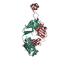
 Homo sapiens (human)
 Mass: 118.174 Da / Num. of mol.: 3 / Source method: obtained synthetically / Formula: C6H14O2 / Comment: precipitant*YM
 Mass: 18.015 Da / Num. of mol.: 66 / Source method: isolated from a natural source / Formula: H2O
 Sample preparation
 Processing