 Movie
Movie Controller


+ Open data

- Basic information
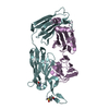
| Entry | Database: PDB / ID: 6e8v | ||||||
|---|---|---|---|---|---|---|---|
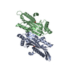
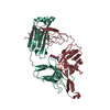
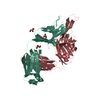
| Title | The crystal structure of bovine ultralong antibody BOV-1 | ||||||
 Components Components |
| ||||||
 Keywords Keywords | IMMUNE SYSTEM / B-lymphocytes / antigen-antibody reactions / antibodies / monoclonal / antibody diversity / Bos taurus | ||||||
| Function / homology |  Function and homology information Function and homology information | ||||||
| Biological species |  | ||||||
| Method |  X-RAY DIFFRACTION / SYNCHROTRON / MOLECULAR REPLACEMENT / Resolution: 3.79 Å X-RAY DIFFRACTION / SYNCHROTRON / MOLECULAR REPLACEMENT / Resolution: 3.79 Å | ||||||
 Authors Authors | Dong, J. / Crowe, J.E. | ||||||
 Citation Citation | Journal: Front Immunol / Year: 2019 Title: Structural Diversity of Ultralong CDRH3s in Seven Bovine Antibody Heavy Chains. Authors: Dong, J. / Finn, J.A. / Larsen, P.A. / Smith, T.P.L. / Crowe Jr., J.E. | ||||||
| History |
|
- Structure visualization
| Structure viewer | Molecule: MolmilJmol/JSmol |
|---|
- Downloads & links
-Download
| PDBx/mmCIF format | 6e8v.cif.gz | 1.2 MB | Display | PDBx/mmCIF format |
|---|---|---|---|---|
| PDB format | pdb6e8v.ent.gz | 1.1 MB | Display | PDB format |
| PDBx/mmJSON format | 6e8v.json.gz | Tree view | PDBx/mmJSON format | |
| Others |  Other downloads Other downloads |
-Validation report
| Arichive directory | https://data.pdbj.org/pub/pdb/validation_reports/e8/6e8vftp://data.pdbj.org/pub/pdb/validation_reports/e8/6e8v | HTTPS FTP |
|---|
-Related structure data
| Related structure data | 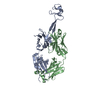 6e9gC 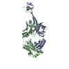 6e9hC 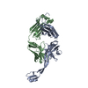 6e9iC 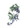 6e9kC 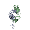 6e9qC  6e9uC C: citing same article ( |
|---|---|
| Similar structure data |


-Links
 PDBj
PDBj

- Assembly
| Deposited unit | 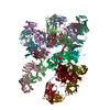
| ||||||||
|---|---|---|---|---|---|---|---|---|---|
| 1 | 
| ||||||||
| 2 | 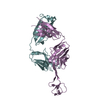
| ||||||||
| 3 | 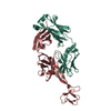
| ||||||||
| 4 | 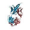
| ||||||||
| 5 | 
| ||||||||
| 6 | 
| ||||||||
| 7 | 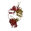
| ||||||||
| 8 | 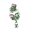
| ||||||||
| Unit cell |
|
-Components
| #1: Antibody | Mass: 28554.646 Da / Num. of mol.: 8 Source method: isolated from a genetically manipulated source Source: (gene. exp.)  Homo sapiens (human) Homo sapiens (human)#2: Antibody | Mass: 22549.596 Da / Num. of mol.: 8 Source method: isolated from a genetically manipulated source Source: (gene. exp.) Homo sapiens (human) / References: UniProt: Q3T101Has protein modification | Y | |
|---|
-Experimental details
-Experiment
| Experiment | Method: X-RAY DIFFRACTION / Number of used crystals: 1 |
|---|
- Sample preparation
| Crystal | Density Matthews: 4.15 Å3/Da / Density % sol: 70.36 % |
|---|---|
| Crystal grow | Temperature: 291 K / Method: evaporation Details: 14-18% PEG 3350, 0.1 M Citric acid pH 4.0-45., 0.1 M sodium citrate tribasic |
-Data collection
| Diffraction | Mean temperature: 100 K / Serial crystal experiment: N |
|---|---|
| Diffraction source | Source: SYNCHROTRON / Site: APS  / Beamline: 21-ID-G / Wavelength: 0.97856 Å / Beamline: 21-ID-G / Wavelength: 0.97856 Å |
| Detector | Type: RAYONIX MX300HE / Detector: CCD / Date: Jul 21, 2016 |
| Radiation | Protocol: SINGLE WAVELENGTH / Monochromatic (M) / Laue (L): M / Scattering type: x-ray |
| Radiation wavelength | Wavelength: 0.97856 Å / Relative weight: 1 |
| Reflection | Resolution: 3.196→49.84 Å / Num. obs: 89101 / % possible obs: 100 % / Redundancy: 7.5 % Data reduction details: The data is highly anisotropic. The diffraction anisotropy server at UCLA was used to anisotropically scale the data to 3.20 angstrom along a*, b* axes, and to 3.80 angstrom along c* axis Rmerge(I) obs: 0.216 / Net I/σ(I): 8.5 |
| Reflection shell | Resolution: 3.196→3.2318 Å / Rmerge(I) obs: 1.44 / Num. unique all: 3534 |
- Processing
| Software |
| ||||||||||||||||||||||||||||||||||||||||||||||||||||||||||||||||||||||||||||||||||||||||||||||||||||||||||||||||||||||||||||||||||||||||||||||||||||||||||||||||||||||||
|---|---|---|---|---|---|---|---|---|---|---|---|---|---|---|---|---|---|---|---|---|---|---|---|---|---|---|---|---|---|---|---|---|---|---|---|---|---|---|---|---|---|---|---|---|---|---|---|---|---|---|---|---|---|---|---|---|---|---|---|---|---|---|---|---|---|---|---|---|---|---|---|---|---|---|---|---|---|---|---|---|---|---|---|---|---|---|---|---|---|---|---|---|---|---|---|---|---|---|---|---|---|---|---|---|---|---|---|---|---|---|---|---|---|---|---|---|---|---|---|---|---|---|---|---|---|---|---|---|---|---|---|---|---|---|---|---|---|---|---|---|---|---|---|---|---|---|---|---|---|---|---|---|---|---|---|---|---|---|---|---|---|---|---|---|---|---|---|---|---|
| Refinement | Method to determine structure: MOLECULAR REPLACEMENT / Resolution: 3.79→49.203 Å / SU ML: 0.47 / Cross valid method: FREE R-VALUE / σ(F): 1.34 / Phase error: 29.11 / Stereochemistry target values: ML
| ||||||||||||||||||||||||||||||||||||||||||||||||||||||||||||||||||||||||||||||||||||||||||||||||||||||||||||||||||||||||||||||||||||||||||||||||||||||||||||||||||||||||
| Solvent computation | Shrinkage radii: 0.9 Å / VDW probe radii: 1.11 Å / Solvent model: FLAT BULK SOLVENT MODEL | ||||||||||||||||||||||||||||||||||||||||||||||||||||||||||||||||||||||||||||||||||||||||||||||||||||||||||||||||||||||||||||||||||||||||||||||||||||||||||||||||||||||||
| Refinement step | Cycle: LAST / Resolution: 3.79→49.203 Å
| ||||||||||||||||||||||||||||||||||||||||||||||||||||||||||||||||||||||||||||||||||||||||||||||||||||||||||||||||||||||||||||||||||||||||||||||||||||||||||||||||||||||||
| Refine LS restraints |
| ||||||||||||||||||||||||||||||||||||||||||||||||||||||||||||||||||||||||||||||||||||||||||||||||||||||||||||||||||||||||||||||||||||||||||||||||||||||||||||||||||||||||
| LS refinement shell |
| ||||||||||||||||||||||||||||||||||||||||||||||||||||||||||||||||||||||||||||||||||||||||||||||||||||||||||||||||||||||||||||||||||||||||||||||||||||||||||||||||||||||||
| Refinement TLS params. | Method: refined / Origin x: 57.6253 Å / Origin y: 209.4699 Å / Origin z: 274.5893 Å
| ||||||||||||||||||||||||||||||||||||||||||||||||||||||||||||||||||||||||||||||||||||||||||||||||||||||||||||||||||||||||||||||||||||||||||||||||||||||||||||||||||||||||
| Refinement TLS group | Selection details: all |
