[English] 日本語
Yorodumi
- PDB-6okq: Crystal structure of the SF12 Fab -

+
Open data


ID or keywords:

Loading...

-
Basic information

Entry
Database: PDB / ID: 6okq
TitleCrystal structure of the SF12 Fab
Components
  • SF12 Fab Heavy Chain,SF12 Fab Heavy Chain
  • SF12 Fab Light Chain,SF12 Fab Light Chain
KeywordsIMMUNE SYSTEM / HIV-1 broadly-neutralizing antibody / Fab structure / silent face / Envelope
Function / homology
Function and homology information


immunoglobulin complex / adaptive immune response / extracellular region / membrane / plasma membrane
Similarity search - Function
Immunoglobulin V-Type / Immunoglobulin V-set domain / Immunoglobulin V-set domain / Immunoglobulin subtype / Immunoglobulin / Immunoglobulin/major histocompatibility complex, conserved site / Immunoglobulins and major histocompatibility complex proteins signature. / Immunoglobulin C-Type / Immunoglobulin C1-set / Immunoglobulin C1-set domain ...Immunoglobulin V-Type / Immunoglobulin V-set domain / Immunoglobulin V-set domain / Immunoglobulin subtype / Immunoglobulin / Immunoglobulin/major histocompatibility complex, conserved site / Immunoglobulins and major histocompatibility complex proteins signature. / Immunoglobulin C-Type / Immunoglobulin C1-set / Immunoglobulin C1-set domain / Ig-like domain profile. / Immunoglobulin-like domain / Immunoglobulin-like domain superfamily / Immunoglobulins / Immunoglobulin-like fold / Immunoglobulin-like / Sandwich / Mainly Beta
Similarity search - Domain/homology
Immunoglobulin gamma-1 heavy chain / Ig-like domain-containing protein
Similarity search - Component
Biological speciesHomo sapiens (human)
MethodX-RAY DIFFRACTION / SYNCHROTRON / MOLECULAR REPLACEMENT / Resolution: 3.2 Å
AuthorsBarnes, C.O. / Bjorkman, P.J.
Funding support United States, 2items
OrganizationGrant numberCountry
National Institutes of Health/National Institute Of Allergy and Infectious Diseases (NIH/NIAID)P01 AI100148 United States
National Institutes of Health/National Institute of General Medical Sciences (NIH/NIGMS)P50 GM082545-06 United States
Citation
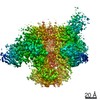
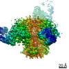
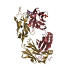
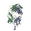
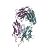
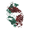
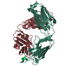
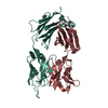
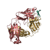
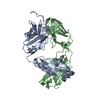
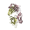
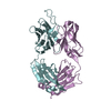
Journal: Immunity / Year: 2019
Title: Broad and Potent Neutralizing Antibodies Recognize the Silent Face of the HIV Envelope.
Authors: Till Schoofs / Christopher O Barnes / Nina Suh-Toma / Jovana Golijanin / Philipp Schommers / Henning Gruell / Anthony P West / Franziska Bach / Yu Erica Lee / Lilian Nogueira / Ivelin S ...Authors: Till Schoofs / Christopher O Barnes / Nina Suh-Toma / Jovana Golijanin / Philipp Schommers / Henning Gruell / Anthony P West / Franziska Bach / Yu Erica Lee / Lilian Nogueira / Ivelin S Georgiev / Robert T Bailer / Julie Czartoski / John R Mascola / Michael S Seaman / M Juliana McElrath / Nicole A Doria-Rose / Florian Klein / Michel C Nussenzweig / Pamela J Bjorkman /
Abstract: Broadly neutralizing antibodies (bNAbs) against HIV-1 envelope (Env) inform vaccine design and are potential therapeutic agents. We identified SF12 and related bNAbs with up to 62% neutralization ...Broadly neutralizing antibodies (bNAbs) against HIV-1 envelope (Env) inform vaccine design and are potential therapeutic agents. We identified SF12 and related bNAbs with up to 62% neutralization breadth from an HIV-infected donor. SF12 recognized a glycan-dominated epitope on Env's silent face and was potent against clade AE viruses, which are poorly covered by V3-glycan bNAbs. A 3.3Å cryo-EM structure of a SF12-Env trimer complex showed additional contacts to Env protein residues by SF12 compared with VRC-PG05, the only other known donor-derived silentface antibody, explaining SF12's increased neutralization breadth, potency, and resistance to Env mutation routes. Asymmetric binding of SF12 was associated with distinct N-glycan conformations across Env protomers, demonstrating intra-Env glycan heterogeneity. Administrating SF12 to HIV-1-infected humanized mice suppressed viremia and selected for viruses lacking the N448 glycan. Effective bNAbs can therefore be raised against HIV-1 Env's silent face, suggesting their potential for HIV-1 prevention, therapy, and vaccine development.
History
DepositionApr 14, 2019Deposition site: RCSB / Processing site: RCSB
Revision 1.0Jun 5, 2019Provider: repository / Type: Initial release
Revision 1.1Jul 3, 2019Group: Data collection / Database references / Category: citation / Item: _citation.journal_volume / _citation.page_first
Revision 1.2Dec 18, 2019Group: Author supporting evidence / Category: pdbx_audit_support / Item: _pdbx_audit_support.funding_organization
Revision 1.3Oct 11, 2023Group: Data collection / Database references / Refinement description
Category: chem_comp_atom / chem_comp_bond ...chem_comp_atom / chem_comp_bond / database_2 / pdbx_initial_refinement_model
Item: _database_2.pdbx_DOI / _database_2.pdbx_database_accession

-
Structure visualization

Structure viewerMolecule:
MolmilJmol/JSmol

Downloads & links

-
Assembly

Deposited unit
A: SF12 Fab Heavy Chain,SF12 Fab Heavy Chain
B: SF12 Fab Light Chain,SF12 Fab Light Chain
C: SF12 Fab Light Chain,SF12 Fab Light Chain
D: SF12 Fab Heavy Chain,SF12 Fab Heavy Chain
E: SF12 Fab Light Chain,SF12 Fab Light Chain
F: SF12 Fab Heavy Chain,SF12 Fab Heavy Chain


Theoretical massNumber of molelcules
Total (without water)147,7936
Polymers147,7936
Non-polymers00
Water0
1
A: SF12 Fab Heavy Chain,SF12 Fab Heavy Chain
B: SF12 Fab Light Chain,SF12 Fab Light Chain


Theoretical massNumber of molelcules
Total (without water)49,2642
Polymers49,2642
Non-polymers00
Water0
TypeNameSymmetry operationNumber
identity operation1_555x,y,z1
Buried area3470 Å2
ΔGint-17 kcal/mol
Surface area19740 Å2
MethodPISA
2
C: SF12 Fab Light Chain,SF12 Fab Light Chain
D: SF12 Fab Heavy Chain,SF12 Fab Heavy Chain


Theoretical massNumber of molelcules
Total (without water)49,2642
Polymers49,2642
Non-polymers00
Water0
TypeNameSymmetry operationNumber
identity operation1_555x,y,z1
Buried area3560 Å2
ΔGint-20 kcal/mol
Surface area19580 Å2
MethodPISA
3
E: SF12 Fab Light Chain,SF12 Fab Light Chain
F: SF12 Fab Heavy Chain,SF12 Fab Heavy Chain


Theoretical massNumber of molelcules
Total (without water)49,2642
Polymers49,2642
Non-polymers00
Water0
TypeNameSymmetry operationNumber
identity operation1_555x,y,z1
Buried area3520 Å2
ΔGint-19 kcal/mol
Surface area19600 Å2
MethodPISA
Unit cell
Length a, b, c (Å)223.018, 223.018, 288.296
Angle α, β, γ (deg.)90.00, 90.00, 120.00
Int Tables number180
Space group name H-MP6222

-
Components

#1: Antibody SF12 Fab Heavy Chain,SF12 Fab Heavy Chain


Mass: 26155.338 Da / Num. of mol.: 3
Source method: isolated from a genetically manipulated source
Source: (gene. exp.) Homo sapiens (human) / Plasmid: pTT5 / Cell line (production host): HEK293 / Production host: Homo sapiens (human) / References: UniProt: P0DOX5
#2: Antibody SF12 Fab Light Chain,SF12 Fab Light Chain


Mass: 23108.842 Da / Num. of mol.: 3
Source method: isolated from a genetically manipulated source
Source: (gene. exp.) Homo sapiens (human) / Plasmid: pTT5 / Cell line (production host): HEK293 / Production host: Homo sapiens (human) / References: UniProt: Q8TCD0

-
Experimental details

-
Experiment

ExperimentMethod: X-RAY DIFFRACTION / Number of used crystals: 1

-
Sample preparation

CrystalDensity Matthews: 7 Å3/Da / Density % sol: 82.43 %
Crystal growTemperature: 293 K / Method: vapor diffusion, hanging drop / pH: 7.5 / Details: 100 mM HEPES, pH 7.5, 1.8 M sodium formate

-
Data collection

DiffractionMean temperature: 100 K / Serial crystal experiment: N
Diffraction sourceSource: SYNCHROTRON / Site: SSRL / Beamline: BL12-2 / Wavelength: 1 Å
DetectorType: DECTRIS PILATUS3 S 6M / Detector: PIXEL / Date: May 19, 2018
RadiationMonochromator: double crystal Si(111) / Protocol: SINGLE WAVELENGTH / Monochromatic (M) / Laue (L): M / Scattering type: x-ray
Radiation wavelengthWavelength: 1 Å / Relative weight: 1
ReflectionResolution: 3.2→39.28 Å / Num. obs: 70865 / % possible obs: 99.6 % / Redundancy: 60.4 % / Biso Wilson estimate: 81.6 Å2 / Rmerge(I) obs: 0.216 / Net I/σ(I): 22.8
Reflection shellResolution: 3.2→3.26 Å / Redundancy: 56.8 % / Rmerge(I) obs: 2.55 / Mean I/σ(I) obs: 1.4 / Num. unique obs: 4473 / % possible all: 98.7

-
Processing

Software
NameVersionClassification
PHENIX(1.14_3219: ???)refinement
XDSdata reduction
Aimlessdata scaling
PHASERphasing
RefinementMethod to determine structure: MOLECULAR REPLACEMENT
Starting model: PDB ENTRY 1U6A
Resolution: 3.2→39.25 Å / SU ML: 0.46 / Cross valid method: FREE R-VALUE / σ(F): 1.33 / Phase error: 40.91
RfactorNum. reflection% reflection
Rfree0.299 3451 4.95 %
Rwork0.273 --
obs0.275 69737 99.6 %
Solvent computationShrinkage radii: 0.9 Å / VDW probe radii: 1.11 Å
Refinement stepCycle: LAST / Resolution: 3.2→39.25 Å
ProteinNucleic acidLigandSolventTotal
Num. atoms10056 0 0 0 10056
Refine LS restraints
Refine-IDTypeDev idealNumber
X-RAY DIFFRACTIONf_bond_d0.0110311
X-RAY DIFFRACTIONf_angle_d1.41414049
X-RAY DIFFRACTIONf_dihedral_angle_d13.7786162
X-RAY DIFFRACTIONf_chiral_restr0.0741581
X-RAY DIFFRACTIONf_plane_restr0.011794
LS refinement shell
Resolution (Å)Rfactor RfreeNum. reflection RfreeRfactor RworkNum. reflection RworkRefine-ID% reflection obs (%)
3.2-3.24380.58311390.55872549X-RAY DIFFRACTION98
3.2438-3.29020.50861180.532587X-RAY DIFFRACTION99
3.2902-3.33920.53231350.49082629X-RAY DIFFRACTION99
3.3392-3.39140.52171380.49112597X-RAY DIFFRACTION100
3.3914-3.4470.50441420.47852598X-RAY DIFFRACTION100
3.447-3.50640.48791270.44442616X-RAY DIFFRACTION99
3.5064-3.57010.48391390.43242607X-RAY DIFFRACTION99
3.5701-3.63870.41811340.382613X-RAY DIFFRACTION100
3.6387-3.71290.40381660.37392613X-RAY DIFFRACTION100
3.7129-3.79360.35871280.34722621X-RAY DIFFRACTION99
3.7936-3.88180.33981290.31592625X-RAY DIFFRACTION100
3.8818-3.97870.29381440.30682639X-RAY DIFFRACTION100
3.9787-4.08620.27831230.27632643X-RAY DIFFRACTION100
4.0862-4.20630.25851520.27172638X-RAY DIFFRACTION100
4.2063-4.34190.2521440.25192627X-RAY DIFFRACTION100
4.3419-4.49690.29691140.21982670X-RAY DIFFRACTION100
4.4969-4.67670.23241360.22232630X-RAY DIFFRACTION99
4.6767-4.88910.27541400.21872657X-RAY DIFFRACTION100
4.8891-5.14640.27631530.22762660X-RAY DIFFRACTION100
5.1464-5.4680.27881510.23372667X-RAY DIFFRACTION100
5.468-5.88890.26411330.23942703X-RAY DIFFRACTION100
5.8889-6.47910.26961360.25052694X-RAY DIFFRACTION100
6.4791-7.41120.28331230.2532737X-RAY DIFFRACTION100
7.4112-9.31670.20991460.21592772X-RAY DIFFRACTION100
9.3167-39.24760.25911610.22692894X-RAY DIFFRACTION99

+
About Yorodumi

-
News

-
Feb 9, 2022. New format data for meta-information of EMDB entries

New format data for meta-information of EMDB entries

  • Version 3 of the EMDB header file is now the official format.
  • The previous official version 1.9 will be removed from the archive.

Related info.:EMDB header

External links:wwPDB to switch to version 3 of the EMDB data model

-
Aug 12, 2020. Covid-19 info

Covid-19 info

URL: https://pdbj.org/emnavi/covid19.php

New page: Covid-19 featured information page in EM Navigator.

Related info.:Covid-19 info / Mar 5, 2020. Novel coronavirus structure data

+
Mar 5, 2020. Novel coronavirus structure data

Novel coronavirus structure data

Related info.:Yorodumi Speices / Aug 12, 2020. Covid-19 info

External links:COVID-19 featured content - PDBj / Molecule of the Month (242):Coronavirus Proteases

+
Jan 31, 2019. EMDB accession codes are about to change! (news from PDBe EMDB page)

EMDB accession codes are about to change! (news from PDBe EMDB page)

  • The allocation of 4 digits for EMDB accession codes will soon come to an end. Whilst these codes will remain in use, new EMDB accession codes will include an additional digit and will expand incrementally as the available range of codes is exhausted. The current 4-digit format prefixed with “EMD-” (i.e. EMD-XXXX) will advance to a 5-digit format (i.e. EMD-XXXXX), and so on. It is currently estimated that the 4-digit codes will be depleted around Spring 2019, at which point the 5-digit format will come into force.
  • The EM Navigator/Yorodumi systems omit the EMD- prefix.

Related info.:Q: What is EMD? / ID/Accession-code notation in Yorodumi/EM Navigator

External links:EMDB Accession Codes are Changing Soon! / Contact to PDBj

+
Jul 12, 2017. Major update of PDB

Major update of PDB

  • wwPDB released updated PDB data conforming to the new PDBx/mmCIF dictionary.
  • This is a major update changing the version number from 4 to 5, and with Remediation, in which all the entries are updated.
  • In this update, many items about electron microscopy experimental information are reorganized (e.g. em_software).
  • Now, EM Navigator and Yorodumi are based on the updated data.

External links:wwPDB Remediation / Enriched Model Files Conforming to OneDep Data Standards Now Available in the PDB FTP Archive

-
Yorodumi

Thousand views of thousand structures

  • Yorodumi is a browser for structure data from EMDB, PDB, SASBDB, etc.
  • This page is also the successor to EM Navigator detail page, and also detail information page/front-end page for Omokage search.
  • The word "yorodu" (or yorozu) is an old Japanese word meaning "ten thousand". "mi" (miru) is to see.

Related info.:EMDB / PDB / SASBDB / Comparison of 3 databanks / Yorodumi Search / Aug 31, 2016. New EM Navigator & Yorodumi / Yorodumi Papers / Jmol/JSmol / Function and homology information / Changes in new EM Navigator and Yorodumi

Read more