 Movie
Movie Controller

[English] 日本語
 Yorodumi
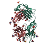
- PDB-6oft: The crystal structure of the first half of the periplasmic protea... -
+ Open data

- Basic information
| Entry | Database: PDB / ID: 6oft | ||||||
|---|---|---|---|---|---|---|---|
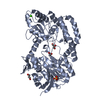
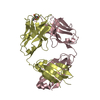
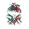
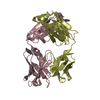
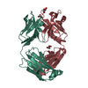
| Title | The crystal structure of the first half of the periplasmic protease PqqL from Escherichia coli | ||||||
 Components Components | Probable zinc protease PqqL | ||||||
 Keywords Keywords | HYDROLASE / Protease / M16 Family / Processing Protease | ||||||
| Function / homology |  Function and homology information Function and homology informationHydrolases; Acting on peptide bonds (peptidases); Metalloendopeptidases / metalloendopeptidase activity / metallopeptidase activity / peptidase activity / outer membrane-bounded periplasmic space / proteolysis / zinc ion binding / cytosol Similarity search - Function | ||||||
| Biological species |  | ||||||
| Method |  X-RAY DIFFRACTION / SYNCHROTRON / MOLECULAR REPLACEMENT / Resolution: 2 Å X-RAY DIFFRACTION / SYNCHROTRON / MOLECULAR REPLACEMENT / Resolution: 2 Å | ||||||
 Authors Authors | Grinter, R. | ||||||
| Funding support |  United Kingdom, 1items United Kingdom, 1items
| ||||||
 Citation Citation | Journal: PLoS Genet / Year: 2019 Title: Protease-associated import systems are widespread in Gram-negative bacteria. Authors: Rhys Grinter / Pok Man Leung / Lakshmi C Wijeyewickrema / Dene Littler / Simone Beckham / Robert N Pike / Daniel Walker / Chris Greening / Trevor Lithgow /  Abstract: Bacteria have evolved sophisticated uptake machineries in order to obtain the nutrients required for growth. Gram-negative plant pathogens of the genus Pectobacterium obtain iron from the protein ...Bacteria have evolved sophisticated uptake machineries in order to obtain the nutrients required for growth. Gram-negative plant pathogens of the genus Pectobacterium obtain iron from the protein ferredoxin, which is produced by their plant hosts. This iron-piracy is mediated by the ferredoxin uptake system (Fus), a gene cluster encoding proteins that transport ferredoxin into the bacterial cell and process it proteolytically. In this work we show that gene clusters related to the Fus are widespread in bacterial species. Through structural and biochemical characterisation of the distantly related Fus homologues YddB and PqqL from Escherichia coli, we show that these proteins are analogous to components of the Fus from Pectobacterium. The membrane protein YddB shares common structural features with the outer membrane ferredoxin transporter FusA, including a large extracellular substrate binding site. PqqL is an active protease with an analogous periplasmic localisation and iron-dependent expression to the ferredoxin processing protease FusC. Structural analysis demonstrates that PqqL and FusC share specific features that distinguish them from other members of the M16 protease family. Taken together, these data provide evidence that protease associated import systems analogous to the Fus are widespread in Gram-negative bacteria. | ||||||
| History |
|
- Structure visualization
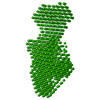
| Structure viewer | Molecule: MolmilJmol/JSmol |
|---|
- Downloads & links
-Download
| PDBx/mmCIF format | 6oft.cif.gz | 392.5 KB | Display | PDBx/mmCIF format |
|---|---|---|---|---|
| PDB format | pdb6oft.ent.gz | 320.4 KB | Display | PDB format |
| PDBx/mmJSON format | 6oft.json.gz | Tree view | PDBx/mmJSON format | |
| Others |  Other downloads Other downloads |
-Validation report
| Arichive directory | https://data.pdbj.org/pub/pdb/validation_reports/of/6oftftp://data.pdbj.org/pub/pdb/validation_reports/of/6oft | HTTPS FTP |
|---|
-Related structure data
| Related structure data |  6ofrC  6ofsC  3amjS S: Starting model for refinement C: citing same article ( |
|---|---|
| Similar structure data |


-Links
 PDBj
PDBj

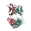
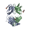
- Assembly
| Deposited unit | 
| ||||||||
|---|---|---|---|---|---|---|---|---|---|
| 1 | 
| ||||||||
| 2 | 
| ||||||||
| Unit cell |
|
-Components
| #1: Protein | Mass: 54179.910 Da / Num. of mol.: 2 Source method: isolated from a genetically manipulated source Source: (gene. exp.) Strain: K12 / Gene: pqqL, yddC, b1494, JW1489 / Production host: References: UniProt: P31828, Hydrolases; Acting on peptide bonds (peptidases); Metalloendopeptidases #2: Chemical |   Mass: 92.094 Da / Num. of mol.: 2 / Source method: obtained synthetically / Formula: C3H8O3 Mass: 92.094 Da / Num. of mol.: 2 / Source method: obtained synthetically / Formula: C3H8O3#3: Chemical |   Mass: 94.971 Da / Num. of mol.: 2 / Source method: obtained synthetically / Formula: PO4 Mass: 94.971 Da / Num. of mol.: 2 / Source method: obtained synthetically / Formula: PO4#4: Chemical |   Mass: 35.453 Da / Num. of mol.: 2 / Source method: obtained synthetically / Formula: Cl Mass: 35.453 Da / Num. of mol.: 2 / Source method: obtained synthetically / Formula: Cl#5: Water | ChemComp-HOH / |  Mass: 18.015 Da / Num. of mol.: 395 / Source method: isolated from a natural source / Formula: H2O Mass: 18.015 Da / Num. of mol.: 395 / Source method: isolated from a natural source / Formula: H2OSequence details | This domain of PqqL was crystallised by in situ Trypsination of the full-length protein. This was ...This domain of PqqL was crystallised by in situ Trypsination of the full-length protein. This was the sequence of the purified protein that was added to the drop (With a small quantity of Trypsin): AALPQDEKLI | |
|---|
-Experimental details
-Experiment
| Experiment | Method: X-RAY DIFFRACTION / Number of used crystals: 1 |
|---|
- Sample preparation
| Crystal | Density Matthews: 2.49 Å3/Da / Density % sol: 50.6 % / Description: Diamond Shaped Plates |
|---|---|
| Crystal grow | Temperature: 293 K / Method: evaporation / pH: 4.2 Details: 0.1 M phosphate-citrate buffer, 0.2 M NaCl, 20% PEG 8000 |
-Data collection
| Diffraction | Mean temperature: 100 K / Serial crystal experiment: N |
|---|---|
| Diffraction source | Source: SYNCHROTRON / Site: Australian Synchrotron / Beamline: MX2 / Wavelength: 0.987 Å |
| Detector | Type: MAR CCD 130 mm / Detector: CCD / Date: Mar 8, 2016 / Details: Yes |
| Radiation | Monochromator: Silicon Crystal / Protocol: SINGLE WAVELENGTH / Monochromatic (M) / Laue (L): M / Scattering type: x-ray |
| Radiation wavelength | Wavelength: 0.987 Å / Relative weight: 1 |
| Reflection | Resolution: 2→21.36 Å / Num. obs: 71853 / % possible obs: 99.8 % / Observed criterion σ(I): 1.7 / Redundancy: 22.3 % / Biso Wilson estimate: 32.01 Å2 / CC1/2: 0.998 / Rmerge(I) obs: 0.348 / Rpim(I) all: 0.07 / Net I/σ(I): 14.4 |
| Reflection shell | Resolution: 2→2.04 Å / Redundancy: 15.1 % / Rmerge(I) obs: 2.009 / Mean I/σ(I) obs: 1.7 / Num. unique obs: 4575 / CC1/2: 0.61 / Rpim(I) all: 0.531 / % possible all: 99 |
- Processing
| Software |
| ||||||||||||||||||||||||||||||||||||||||||||||||||||||||||||||||||||||||||||||||||||||||||||||||||||||||||||||||||
|---|---|---|---|---|---|---|---|---|---|---|---|---|---|---|---|---|---|---|---|---|---|---|---|---|---|---|---|---|---|---|---|---|---|---|---|---|---|---|---|---|---|---|---|---|---|---|---|---|---|---|---|---|---|---|---|---|---|---|---|---|---|---|---|---|---|---|---|---|---|---|---|---|---|---|---|---|---|---|---|---|---|---|---|---|---|---|---|---|---|---|---|---|---|---|---|---|---|---|---|---|---|---|---|---|---|---|---|---|---|---|---|---|---|---|---|
| Refinement | Method to determine structure: MOLECULAR REPLACEMENT Starting model: 3AMJ Resolution: 2→21.36 Å / Cor.coef. Fo:Fc: 0.942 / Cor.coef. Fo:Fc free: 0.919 / SU R Cruickshank DPI: 0.173 / Cross valid method: THROUGHOUT / σ(F): 0 / SU R Blow DPI: 0.171 / SU Rfree Blow DPI: 0.15 / SU Rfree Cruickshank DPI: 0.152
| ||||||||||||||||||||||||||||||||||||||||||||||||||||||||||||||||||||||||||||||||||||||||||||||||||||||||||||||||||
| Displacement parameters | Biso mean: 37.35 Å2
| ||||||||||||||||||||||||||||||||||||||||||||||||||||||||||||||||||||||||||||||||||||||||||||||||||||||||||||||||||
| Refine analyze | Luzzati coordinate error obs: 0.27 Å | ||||||||||||||||||||||||||||||||||||||||||||||||||||||||||||||||||||||||||||||||||||||||||||||||||||||||||||||||||
| Refinement step | Cycle: 1 / Resolution: 2→21.36 Å
| ||||||||||||||||||||||||||||||||||||||||||||||||||||||||||||||||||||||||||||||||||||||||||||||||||||||||||||||||||
| Refine LS restraints |
| ||||||||||||||||||||||||||||||||||||||||||||||||||||||||||||||||||||||||||||||||||||||||||||||||||||||||||||||||||
| LS refinement shell | Resolution: 2→2.01 Å / Total num. of bins used: 50
| ||||||||||||||||||||||||||||||||||||||||||||||||||||||||||||||||||||||||||||||||||||||||||||||||||||||||||||||||||
| Refinement TLS params. | Method: refined / Refine-ID: X-RAY DIFFRACTION
| ||||||||||||||||||||||||||||||||||||||||||||||||||||||||||||||||||||||||||||||||||||||||||||||||||||||||||||||||||
| Refinement TLS group |
|
