 Movie
Movie Controller

[English] 日本語
 Yorodumi
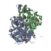
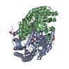
- PDB-6ofr: The crystal structure of the outer membrane transporter YddB from... -
+ Open data

- Basic information
| Entry | Database: PDB / ID: 6ofr | ||||||
|---|---|---|---|---|---|---|---|
| Title | The crystal structure of the outer membrane transporter YddB from Escherichia coli | ||||||
 Components Components | TonB-dependent outer membrane receptor | ||||||
 Keywords Keywords | TRANSPORT PROTEIN / Protein Transport / Gram-negative bacteria / Outer membrane / Nutrient Uptake / TonB-Dependent Transporter | ||||||
| Function / homology | TonB-dependent receptor (TBDR) proteins profile. / Vitamin B12 transporter BtuB-like / TonB-dependent receptor, plug domain superfamily / TonB-dependent receptor, plug domain / TonB-dependent Receptor Plug Domain / TonB-dependent receptor-like, beta-barrel domain superfamily / cell outer membrane / TonB-dependent receptor / Uncharacterized protein YddB Function and homology information Function and homology information | ||||||
| Biological species |  | ||||||
| Method |  X-RAY DIFFRACTION / SYNCHROTRON / MOLECULAR REPLACEMENT / Resolution: 2.4 Å X-RAY DIFFRACTION / SYNCHROTRON / MOLECULAR REPLACEMENT / Resolution: 2.4 Å | ||||||
 Authors Authors | Grinter, R. | ||||||
| Funding support |  United Kingdom, 1items United Kingdom, 1items
| ||||||
 Citation Citation | Journal: PLoS Genet / Year: 2019 Title: Protease-associated import systems are widespread in Gram-negative bacteria. Authors: Rhys Grinter / Pok Man Leung / Lakshmi C Wijeyewickrema / Dene Littler / Simone Beckham / Robert N Pike / Daniel Walker / Chris Greening / Trevor Lithgow /  Abstract: Bacteria have evolved sophisticated uptake machineries in order to obtain the nutrients required for growth. Gram-negative plant pathogens of the genus Pectobacterium obtain iron from the protein ...Bacteria have evolved sophisticated uptake machineries in order to obtain the nutrients required for growth. Gram-negative plant pathogens of the genus Pectobacterium obtain iron from the protein ferredoxin, which is produced by their plant hosts. This iron-piracy is mediated by the ferredoxin uptake system (Fus), a gene cluster encoding proteins that transport ferredoxin into the bacterial cell and process it proteolytically. In this work we show that gene clusters related to the Fus are widespread in bacterial species. Through structural and biochemical characterisation of the distantly related Fus homologues YddB and PqqL from Escherichia coli, we show that these proteins are analogous to components of the Fus from Pectobacterium. The membrane protein YddB shares common structural features with the outer membrane ferredoxin transporter FusA, including a large extracellular substrate binding site. PqqL is an active protease with an analogous periplasmic localisation and iron-dependent expression to the ferredoxin processing protease FusC. Structural analysis demonstrates that PqqL and FusC share specific features that distinguish them from other members of the M16 protease family. Taken together, these data provide evidence that protease associated import systems analogous to the Fus are widespread in Gram-negative bacteria. | ||||||
| History |
|
- Structure visualization
| Structure viewer | Molecule: MolmilJmol/JSmol |
|---|
- Downloads & links
-Download
| PDBx/mmCIF format | 6ofr.cif.gz | 330.6 KB | Display | PDBx/mmCIF format |
|---|---|---|---|---|
| PDB format | pdb6ofr.ent.gz | 266.3 KB | Display | PDB format |
| PDBx/mmJSON format | 6ofr.json.gz | Tree view | PDBx/mmJSON format | |
| Others |  Other downloads Other downloads |
-Validation report
| Arichive directory | https://data.pdbj.org/pub/pdb/validation_reports/of/6ofrftp://data.pdbj.org/pub/pdb/validation_reports/of/6ofr | HTTPS FTP |
|---|
-Related structure data
| Related structure data |  6ofsC 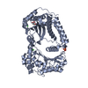 6oftC 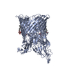 4zgvS S: Starting model for refinement C: citing same article ( |
|---|---|
| Similar structure data |

-Links
 PDBj
PDBj- Assembly
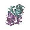
| Deposited unit | 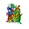
| ||||||||
|---|---|---|---|---|---|---|---|---|---|
| 1 |
| ||||||||
| Unit cell |
|
-Components
| #1: Protein | Mass: 86302.805 Da / Num. of mol.: 1 Source method: isolated from a genetically manipulated source Source: (gene. exp.) Gene: yddB, ACU57_09575, BANRA_00721, BANRA_04049, BvCmsKSP026_02313, BvCmsSINP012_01435, C5N07_00015, CA593_20420, D0X26_01860, D3821_23605, DNQ41_11905, E4Z89_11620, E5M00_15980, EAI52_23140, ...Gene: yddB, ACU57_09575, BANRA_00721, BANRA_04049, BvCmsKSP026_02313, BvCmsSINP012_01435, C5N07_00015, CA593_20420, D0X26_01860, D3821_23605, DNQ41_11905, E4Z89_11620, E5M00_15980, EAI52_23140, EC3234A_33c00020, EC3426_02521, EPS71_22575, EXX71_23015, EYD11_11385, NCTC13462_06167, NCTC9037_02822, NCTC9045_02987, NCTC9062_00632, NCTC9073_01172, RK56_010145, SAMEA3753300_05217 Production host: | ||||||||||
|---|---|---|---|---|---|---|---|---|---|---|---|
| #2: Sugar | ChemComp-BOG /   Type: D-saccharide / Mass: 292.369 Da / Num. of mol.: 7 Type: D-saccharide / Mass: 292.369 Da / Num. of mol.: 7Source method: isolated from a genetically manipulated source Formula: C14H28O6 / Comment: detergent*YM #3: Chemical |   Mass: 24.305 Da / Num. of mol.: 2 / Source method: obtained synthetically / Formula: Mg Mass: 24.305 Da / Num. of mol.: 2 / Source method: obtained synthetically / Formula: Mg#4: Chemical |   Mass: 92.094 Da / Num. of mol.: 3 / Source method: obtained synthetically / Formula: C3H8O3 Mass: 92.094 Da / Num. of mol.: 3 / Source method: obtained synthetically / Formula: C3H8O3#5: Water | ChemComp-HOH / |  Mass: 18.015 Da / Num. of mol.: 244 / Source method: isolated from a natural source / Formula: H2O Mass: 18.015 Da / Num. of mol.: 244 / Source method: isolated from a natural source / Formula: H2OHas ligand of interest | N | Has protein modification | Y | |
-Experimental details
-Experiment
| Experiment | Method: X-RAY DIFFRACTION / Number of used crystals: 1 |
|---|
- Sample preparation
| Crystal | Density Matthews: 3.52 Å3/Da / Density % sol: 65.02 % |
|---|---|
| Crystal grow | Temperature: 293 K / Method: evaporation / pH: 6.5 Details: 0.1 M Na cacodylate, 0.15 M Ca acetate, 15 % PEG 8000 and 20 % glycerol |
-Data collection
| Diffraction | Mean temperature: 100 K / Serial crystal experiment: N |
|---|---|
| Diffraction source | Source: SYNCHROTRON / Site: Australian Synchrotron / Beamline: MX2 / Wavelength: 0.987 Å |
| Detector | Type: MARMOSAIC 300 mm CCD / Detector: CCD / Date: Sep 11, 2015 / Details: Yes |
| Radiation | Monochromator: Silicon Crystal / Protocol: SINGLE WAVELENGTH / Monochromatic (M) / Laue (L): M / Scattering type: x-ray |
| Radiation wavelength | Wavelength: 0.987 Å / Relative weight: 1 |
| Reflection | Resolution: 2.4→47.92 Å / Num. obs: 49176 / % possible obs: 99.6 % / Redundancy: 10.4 % / Biso Wilson estimate: 56.31 Å2 / CC1/2: 0.991 / Rmerge(I) obs: 0.219 / Rpim(I) all: 0.072 / Net I/σ(I): 6 |
| Reflection shell | Resolution: 2.4→2.48 Å / Redundancy: 10.7 % / Rmerge(I) obs: 1.96 / Mean I/σ(I) obs: 1.5 / Num. unique obs: 4454 / CC1/2: 0.722 / Rpim(I) all: 0.622 / % possible all: 99.9 |
- Processing
| Software |
| |||||||||||||||
|---|---|---|---|---|---|---|---|---|---|---|---|---|---|---|---|---|
| Refinement | Method to determine structure: MOLECULAR REPLACEMENT Starting model: 4ZGV Resolution: 2.4→47.92 Å / Cross valid method: FREE R-VALUE /
| |||||||||||||||
| Refinement step | Cycle: LAST / Resolution: 2.4→47.92 Å
|
