 Movie
Movie Controller

[English] 日本語
 Yorodumi
- PDB-4zgv: The Crystal Structure of the Ferredoxin Receptor FusA from Pectob... -
+ Open data

- Basic information
| Entry | Database: PDB / ID: 4zgv | ||||||
|---|---|---|---|---|---|---|---|
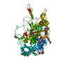
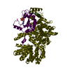
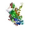
| Title | The Crystal Structure of the Ferredoxin Receptor FusA from Pectobacterium atrosepticum SCRI1043 | ||||||
 Components Components | Ferredoxin receptor | ||||||
 Keywords Keywords | TRANSPORT PROTEIN / Beta-barrel / TonB-dependent receptor / Iron-transporter / Outer membrane | ||||||
| Function / homology | TonB-dependent receptor, plug domain superfamily / TonB-dependent receptor, plug domain / TonB-dependent Receptor Plug Domain / TonB-dependent receptor-like, beta-barrel domain superfamily / cell outer membrane / TonB-dependent receptor plug domain-containing protein Function and homology information Function and homology information | ||||||
| Biological species |  Pectobacterium atrosepticum (bacteria) Pectobacterium atrosepticum (bacteria) | ||||||
| Method |  X-RAY DIFFRACTION / SYNCHROTRON / SAD / Resolution: 3.2 Å X-RAY DIFFRACTION / SYNCHROTRON / SAD / Resolution: 3.2 Å | ||||||
 Authors Authors | Grinter, R. / Josts, I. / Roszak, A.W. / Cogdell, R.J. / Walker, D. | ||||||
 Citation Citation | Journal: Nat Commun / Year: 2016 Title: Structure of the bacterial plant-ferredoxin receptor FusA. Authors: Grinter, R. / Josts, I. / Mosbahi, K. / Roszak, A.W. / Cogdell, R.J. / Bonvin, A.M. / Milner, J.J. / Kelly, S.M. / Byron, O. / Smith, B.O. / Walker, D. | ||||||
| History |
|
- Structure visualization
| Structure viewer | Molecule: MolmilJmol/JSmol |
|---|
- Downloads & links
-Download
| PDBx/mmCIF format | 4zgv.cif.gz | 657 KB | Display | PDBx/mmCIF format |
|---|---|---|---|---|
| PDB format | pdb4zgv.ent.gz | 542.9 KB | Display | PDB format |
| PDBx/mmJSON format | 4zgv.json.gz | Tree view | PDBx/mmJSON format | |
| Others |  Other downloads Other downloads |
-Validation report
| Arichive directory | https://data.pdbj.org/pub/pdb/validation_reports/zg/4zgvftp://data.pdbj.org/pub/pdb/validation_reports/zg/4zgv | HTTPS FTP |
|---|
-Related structure data


-Links
 PDBj
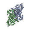
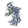
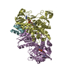
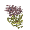
PDBj- Assembly
| Deposited unit | 
| ||||||||
|---|---|---|---|---|---|---|---|---|---|
| 1 | 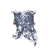
| ||||||||
| 2 | 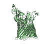
| ||||||||
| Unit cell |
|
-Components
| #1: Protein | Mass: 97786.562 Da / Num. of mol.: 2 Source method: isolated from a genetically manipulated source Source: (gene. exp.) Pectobacterium atrosepticum (bacteria) / Strain: SCRI 1043 / ATCC BAA-672 / Gene: ECA0878 / Plasmid: pETFA1043 / Production host: #2: Chemical |   Mass: 229.402 Da / Num. of mol.: 2 / Source method: obtained synthetically / Formula: C14H31NO / Comment: LDAO, detergent*YM Mass: 229.402 Da / Num. of mol.: 2 / Source method: obtained synthetically / Formula: C14H31NO / Comment: LDAO, detergent*YM#3: Sugar | ChemComp-BOG /   Type: D-saccharide / Mass: 292.369 Da / Num. of mol.: 4 Type: D-saccharide / Mass: 292.369 Da / Num. of mol.: 4Source method: isolated from a genetically manipulated source Formula: C14H28O6 / Comment: detergent*YM #4: Water | ChemComp-HOH / |  Mass: 18.015 Da / Num. of mol.: 2 / Source method: isolated from a natural source / Formula: H2O Mass: 18.015 Da / Num. of mol.: 2 / Source method: isolated from a natural source / Formula: H2OHas protein modification | Y | |
|---|
-Experimental details
-Experiment
| Experiment | Method: X-RAY DIFFRACTION |
|---|
- Sample preparation
| Crystal | Density Matthews: 3.87 Å3/Da / Density % sol: 68.23 % Description: Long thin needles, up to 1 mM by 50 uM. Very fragile, bent easily, difficult to handle |
|---|---|
| Crystal grow | Temperature: 289 K / Method: vapor diffusion, sitting drop / pH: 7.5 Details: 11-14 % PVP, 14 % PEG 2000 MME, 0.1 Tris, 0.05 M MgCl2 pH 7.5, with a FusA concentration of 15 mg.ml-1 in 50 mM Tris, 200 mM NaCl, 0.8-1 % (v/v) beta-OG, 0.4 % LDAO pH 7.9, at 289 K |
-Data collection
| Diffraction | Mean temperature: 100 K |
|---|---|
| Diffraction source | Source: SYNCHROTRON / Site: Diamond  / Beamline: I02 / Wavelength: 0.9765 Å / Beamline: I02 / Wavelength: 0.9765 Å |
| Detector | Type: PSI PILATUS 6M / Detector: PIXEL / Date: Jul 7, 2014 |
| Radiation | Monochromator: Silicon crystal / Protocol: SINGLE WAVELENGTH / Monochromatic (M) / Laue (L): M / Scattering type: x-ray |
| Radiation wavelength | Wavelength: 0.9765 Å / Relative weight: 1 |
| Reflection | Resolution: 3.2→48.95 Å / Num. obs: 49734 / % possible obs: 100 % / Redundancy: 6.6 % / Rmerge(I) obs: 0.305 / Net I/σ(I): 7 |
| Reflection shell | Resolution: 3.2→3.31 Å / Redundancy: 6.4 % / Rmerge(I) obs: 1.73 / Mean I/σ(I) obs: 1.3 / % possible all: 100 |
- Processing
| Software |
| ||||||||||||||||||||||||||||||||||||||||||||||||||||||||||||||||||||||||||||||||||||||||||||||||||||||||||||||||||||||||||||||||||||||||||||||||||||||||||||||||||||||||||||||||||||||
|---|---|---|---|---|---|---|---|---|---|---|---|---|---|---|---|---|---|---|---|---|---|---|---|---|---|---|---|---|---|---|---|---|---|---|---|---|---|---|---|---|---|---|---|---|---|---|---|---|---|---|---|---|---|---|---|---|---|---|---|---|---|---|---|---|---|---|---|---|---|---|---|---|---|---|---|---|---|---|---|---|---|---|---|---|---|---|---|---|---|---|---|---|---|---|---|---|---|---|---|---|---|---|---|---|---|---|---|---|---|---|---|---|---|---|---|---|---|---|---|---|---|---|---|---|---|---|---|---|---|---|---|---|---|---|---|---|---|---|---|---|---|---|---|---|---|---|---|---|---|---|---|---|---|---|---|---|---|---|---|---|---|---|---|---|---|---|---|---|---|---|---|---|---|---|---|---|---|---|---|---|---|---|---|
| Refinement | Method to determine structure: SAD / Resolution: 3.2→48.95 Å / Cor.coef. Fo:Fc: 0.889 / Cor.coef. Fo:Fc free: 0.881 / SU B: 58.57 / SU ML: 0.408 / Cross valid method: THROUGHOUT / ESU R Free: 0.464 / Stereochemistry target values: MAXIMUM LIKELIHOOD / Details: HYDROGENS HAVE BEEN ADDED IN THE RIDING POSITIONS
| ||||||||||||||||||||||||||||||||||||||||||||||||||||||||||||||||||||||||||||||||||||||||||||||||||||||||||||||||||||||||||||||||||||||||||||||||||||||||||||||||||||||||||||||||||||||
| Solvent computation | Ion probe radii: 0.8 Å / Shrinkage radii: 0.8 Å / VDW probe radii: 1.2 Å / Solvent model: MASK | ||||||||||||||||||||||||||||||||||||||||||||||||||||||||||||||||||||||||||||||||||||||||||||||||||||||||||||||||||||||||||||||||||||||||||||||||||||||||||||||||||||||||||||||||||||||
| Displacement parameters | Biso mean: 81.354 Å2
| ||||||||||||||||||||||||||||||||||||||||||||||||||||||||||||||||||||||||||||||||||||||||||||||||||||||||||||||||||||||||||||||||||||||||||||||||||||||||||||||||||||||||||||||||||||||
| Refinement step | Cycle: LAST / Resolution: 3.2→48.95 Å
| ||||||||||||||||||||||||||||||||||||||||||||||||||||||||||||||||||||||||||||||||||||||||||||||||||||||||||||||||||||||||||||||||||||||||||||||||||||||||||||||||||||||||||||||||||||||
| Refine LS restraints |
|