 Movie
Movie Controller


+ Open data

- Basic information
| Entry | Database: PDB / ID: 1rky | |||||||||
|---|---|---|---|---|---|---|---|---|---|---|
| Title | PPLO + Xe | |||||||||
 Components Components | lysyl oxidase | |||||||||
 Keywords Keywords | OXIDOREDUCTASE / PPLO / lysyl oxidase / CAO / CuAO / copper-containing / amine oxidase / oxygen binding site / dioxygen binding site / xenon / TPQ / quinone / trihydroxyphenylalanine quinone | |||||||||
| Function / homology |  Function and homology information Function and homology informationOxidoreductases; Acting on the CH-NH2 group of donors; With oxygen as acceptor / diamine oxidase activity / primary methylamine oxidase activity / amine metabolic process / quinone binding / copper ion binding / plasma membrane Similarity search - Function | |||||||||
| Biological species |  Pichia pastoris (fungus) Pichia pastoris (fungus) | |||||||||
| Method |  X-RAY DIFFRACTION / SYNCHROTRON / MOLECULAR REPLACEMENT / Resolution: 1.68 Å X-RAY DIFFRACTION / SYNCHROTRON / MOLECULAR REPLACEMENT / Resolution: 1.68 Å | |||||||||
 Authors Authors | Guss, J.M. / Duff, A.P. | |||||||||
 Citation Citation | Journal: J.Mol.Biol. / Year: 2004 Title: Using Xenon as a Probe for Dioxygen-binding Sites in Copper Amine Oxidases Authors: Duff, A.P. / Trambaiolo, D.M. / Cohen, A.E. / Ellis, P.J. / Juda, G.A. / Shepard, E.M. / Langley, D.B. / Dooley, D.M. / Freeman, H.C. / Guss, J.M. | |||||||||
| History |
|
- Structure visualization
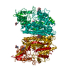
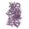
| Structure viewer | Molecule: MolmilJmol/JSmol |
|---|
- Downloads & links
-Download
| PDBx/mmCIF format | 1rky.cif.gz | 188.2 KB | Display | PDBx/mmCIF format |
|---|---|---|---|---|
| PDB format | pdb1rky.ent.gz | 144.9 KB | Display | PDB format |
| PDBx/mmJSON format | 1rky.json.gz | Tree view | PDBx/mmJSON format | |
| Others |  Other downloads Other downloads |
-Validation report
| Arichive directory | https://data.pdbj.org/pub/pdb/validation_reports/rk/1rkyftp://data.pdbj.org/pub/pdb/validation_reports/rk/1rky | HTTPS FTP |
|---|
-Related structure data
| Related structure data | 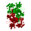 1rjoC  1w2zC 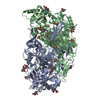 1n9eS S: Starting model for refinement C: citing same article ( |
|---|---|
| Similar structure data |


-Links
 PDBj
PDBj


- Assembly
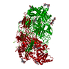
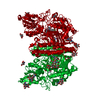
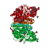
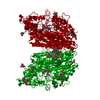
| Deposited unit | 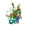
| ||||||||
|---|---|---|---|---|---|---|---|---|---|
| 1 | 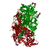
| ||||||||
| Unit cell |
| ||||||||
| Components on special symmetry positions |
| ||||||||
| Details | The biological assembly is a dimer obtained by applying crystallographic symmetry to the monomer. |
-Components
-Protein , 1 types, 1 molecules A
| #1: Protein | Mass: 85289.125 Da / Num. of mol.: 1 / Fragment: RESIDUES 41-787 Source method: isolated from a genetically manipulated source Source: (gene. exp.) Pichia pastoris (fungus) / Gene: ATCC 28, 485 / Plasmid: pPIC3 / Production host: Pichia pastoris (fungus) / Strain (production host): GS115References: GenBank: 13936870, UniProt: Q96X16*PLUS, protein-lysine 6-oxidase |
|---|
-Sugars , 2 types, 5 molecules 
| #2: Polysaccharide | beta-D-mannopyranose-(1-4)-2-acetamido-2-deoxy-beta-D-glucopyranose-(1-4)-2-acetamido-2-deoxy-beta- ...beta-D-mannopyranose-(1-4)-2-acetamido-2-deoxy-beta-D-glucopyranose-(1-4)-2-acetamido-2-deoxy-beta-D-glucopyranose Source method: isolated from a genetically manipulated source |
|---|---|
| #3: Sugar | ChemComp-NAG /  Type: D-saccharide, beta linking / Mass: 221.208 Da / Num. of mol.: 4 Type: D-saccharide, beta linking / Mass: 221.208 Da / Num. of mol.: 4Source method: isolated from a genetically manipulated source Formula: C8H15NO6 |
-Non-polymers , 7 types, 653 molecules 


| #4: Chemical | ChemComp-CU /  Mass: 63.546 Da / Num. of mol.: 1 / Source method: obtained synthetically / Formula: Cu Mass: 63.546 Da / Num. of mol.: 1 / Source method: obtained synthetically / Formula: Cu | ||||||||||
|---|---|---|---|---|---|---|---|---|---|---|---|
| #5: Chemical |  Mass: 40.078 Da / Num. of mol.: 2 / Source method: obtained synthetically / Formula: Ca Mass: 40.078 Da / Num. of mol.: 2 / Source method: obtained synthetically / Formula: Ca#6: Chemical |  Mass: 24.305 Da / Num. of mol.: 3 / Source method: obtained synthetically / Formula: Mg Mass: 24.305 Da / Num. of mol.: 3 / Source method: obtained synthetically / Formula: Mg#7: Chemical | ChemComp-CL /  Mass: 35.453 Da / Num. of mol.: 6 / Source method: obtained synthetically / Formula: Cl Mass: 35.453 Da / Num. of mol.: 6 / Source method: obtained synthetically / Formula: Cl#8: Chemical | ChemComp-IMD / |  Mass: 69.085 Da / Num. of mol.: 1 / Source method: obtained synthetically / Formula: C3H5N2 Mass: 69.085 Da / Num. of mol.: 1 / Source method: obtained synthetically / Formula: C3H5N2#9: Chemical | ChemComp-XE /  Mass: 131.293 Da / Num. of mol.: 8 / Source method: obtained synthetically / Formula: Xe Mass: 131.293 Da / Num. of mol.: 8 / Source method: obtained synthetically / Formula: Xe#10: Water | ChemComp-HOH / | Mass: 18.015 Da / Num. of mol.: 632 / Source method: isolated from a natural source / Formula: H2O |
-Experimental details
-Experiment
| Experiment | Method: X-RAY DIFFRACTION / Number of used crystals: 1 |
|---|
- Sample preparation
| Crystal | Density Matthews: 2.54 Å3/Da / Density % sol: 51.26 % |
|---|---|
| Crystal grow | Temperature: 300 K / Method: vapor diffusion, hanging drop / pH: 8 Details: 35% MPD, Imidazole pH 8.0, 200mM MgCl2, VAPOR DIFFUSION, HANGING DROP, temperature 300K. CRYSTAL EXPOSED TO XENON AT 300 PSI FOR 90 SECONDS THEN FROZEN |
-Data collection
| Diffraction | Mean temperature: 100 K |
|---|---|
| Diffraction source | Source: SYNCHROTRON / Site: SSRL  / Beamline: BL9-2 / Wavelength: 1.552 Å / Beamline: BL9-2 / Wavelength: 1.552 Å |
| Detector | Type: ADSC QUANTUM 315 / Detector: CCD / Date: Apr 1, 2003 / Details: double crystal monochromator |
| Radiation | Monochromator: double crystal monochromator / Protocol: SINGLE WAVELENGTH / Monochromatic (M) / Laue (L): M / Scattering type: x-ray |
| Radiation wavelength | Wavelength: 1.552 Å / Relative weight: 1 |
| Reflection | Resolution: 1.68→31.46 Å / Num. obs: 99719 / % possible obs: 87.7 % / Biso Wilson estimate: 27 Å2 / Rmerge(I) obs: 0.09 / Net I/σ(I): 11.9 |
| Reflection shell | Resolution: 1.68→1.74 Å / Mean I/σ(I) obs: 2.1 / % possible all: 84.6 |
- Processing
| Software |
| ||||||||||||||||||||||||||||||||||||||||||||||||||||||||||||||||||||||||||||||||||||||||||||||||||||||||||||||||||||||||||||||||||||||||||||||||||||||||||||||||
|---|---|---|---|---|---|---|---|---|---|---|---|---|---|---|---|---|---|---|---|---|---|---|---|---|---|---|---|---|---|---|---|---|---|---|---|---|---|---|---|---|---|---|---|---|---|---|---|---|---|---|---|---|---|---|---|---|---|---|---|---|---|---|---|---|---|---|---|---|---|---|---|---|---|---|---|---|---|---|---|---|---|---|---|---|---|---|---|---|---|---|---|---|---|---|---|---|---|---|---|---|---|---|---|---|---|---|---|---|---|---|---|---|---|---|---|---|---|---|---|---|---|---|---|---|---|---|---|---|---|---|---|---|---|---|---|---|---|---|---|---|---|---|---|---|---|---|---|---|---|---|---|---|---|---|---|---|---|---|---|---|---|
| Refinement | Method to determine structure: MOLECULAR REPLACEMENT Starting model: PPLO 1N9E chain A Resolution: 1.68→31.47 Å / Cor.coef. Fo:Fc: 0.973 / Cor.coef. Fo:Fc free: 0.961 / SU B: 1.888 / SU ML: 0.061 / Cross valid method: THROUGHOUT / ESU R: 0.091 / ESU R Free: 0.091 / Stereochemistry target values: MAXIMUM LIKELIHOOD Details: HYDROGENS HAVE BEEN ADDED IN THE RIDING POSITIONS. REFMAC MATRIX WEIGHT = 0.3, BFACTOR WIEGHT = 0.
| ||||||||||||||||||||||||||||||||||||||||||||||||||||||||||||||||||||||||||||||||||||||||||||||||||||||||||||||||||||||||||||||||||||||||||||||||||||||||||||||||
| Solvent computation | Ion probe radii: 0.8 Å / Shrinkage radii: 0.8 Å / VDW probe radii: 1.4 Å / Solvent model: BABINET MODEL WITH MASK | ||||||||||||||||||||||||||||||||||||||||||||||||||||||||||||||||||||||||||||||||||||||||||||||||||||||||||||||||||||||||||||||||||||||||||||||||||||||||||||||||
| Displacement parameters | Biso mean: 33.28 Å2
| ||||||||||||||||||||||||||||||||||||||||||||||||||||||||||||||||||||||||||||||||||||||||||||||||||||||||||||||||||||||||||||||||||||||||||||||||||||||||||||||||
| Refinement step | Cycle: LAST / Resolution: 1.68→31.47 Å
| ||||||||||||||||||||||||||||||||||||||||||||||||||||||||||||||||||||||||||||||||||||||||||||||||||||||||||||||||||||||||||||||||||||||||||||||||||||||||||||||||
| Refine LS restraints |
| ||||||||||||||||||||||||||||||||||||||||||||||||||||||||||||||||||||||||||||||||||||||||||||||||||||||||||||||||||||||||||||||||||||||||||||||||||||||||||||||||
| LS refinement shell | Resolution: 1.68→1.724 Å / Total num. of bins used: 20 /
|
