 Movie
Movie Controller

[English] 日本語
 Yorodumi
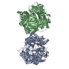
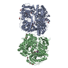
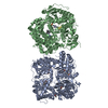
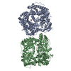
- PDB-3amj: The crystal structure of the heterodimer of M16B peptidase from S... -
+ Open data

- Basic information
| Entry | Database: PDB / ID: 3amj | ||||||
|---|---|---|---|---|---|---|---|
| Title | The crystal structure of the heterodimer of M16B peptidase from Sphingomonas sp. A1 | ||||||
 Components Components |
| ||||||
 Keywords Keywords | HYDROLASE / alpha/beta / zinc peptidase / zinc binding | ||||||
| Function / homology |  Function and homology information Function and homology informationHydrolases; Acting on peptide bonds (peptidases); Metalloendopeptidases / metallopeptidase activity / metal ion binding Similarity search - Function | ||||||
| Biological species |  Sphingomonas (bacteria) Sphingomonas (bacteria) | ||||||
| Method |  X-RAY DIFFRACTION / SYNCHROTRON / MOLECULAR REPLACEMENT / Resolution: 3 Å X-RAY DIFFRACTION / SYNCHROTRON / MOLECULAR REPLACEMENT / Resolution: 3 Å | ||||||
 Authors Authors | Maruyama, Y. / Chuma, A. / Mikami, B. / Hashimoto, W. / Murata, K. | ||||||
 Citation Citation | Journal: J.Mol.Biol. / Year: 2011 Title: Heterosubunit composition and crystal structures of a novel bacterial M16B metallopeptidase Authors: Maruyama, Y. / Chuma, A. / Mikami, B. / Hashimoto, W. / Murata, K. | ||||||
| History |
|
- Structure visualization
| Structure viewer | Molecule: MolmilJmol/JSmol |
|---|
- Downloads & links
-Download
| PDBx/mmCIF format | 3amj.cif.gz | 626.6 KB | Display | PDBx/mmCIF format |
|---|---|---|---|---|
| PDB format | pdb3amj.ent.gz | 514.4 KB | Display | PDB format |
| PDBx/mmJSON format | 3amj.json.gz | Tree view | PDBx/mmJSON format | |
| Others |  Other downloads Other downloads |
-Validation report
| Arichive directory | https://data.pdbj.org/pub/pdb/validation_reports/am/3amjftp://data.pdbj.org/pub/pdb/validation_reports/am/3amj | HTTPS FTP |
|---|
-Related structure data
| Related structure data |  3amiC  3gwdS C: citing same article ( S: Starting model for refinement |
|---|---|
| Similar structure data |


-Links
 PDBj
PDBj

- Assembly
| Deposited unit | 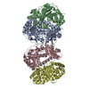
| ||||||||
|---|---|---|---|---|---|---|---|---|---|
| 1 |
| ||||||||
| 2 | 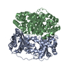
| ||||||||
| 3 | 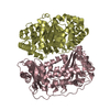
| ||||||||
| Unit cell |
| ||||||||
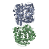
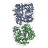
| Details | AUTHOR DEFINED BIOLOGICAL UNIT: UNKNOWN. AUTHOR STATED THAT GEL FILTRATION COLUMN CHROMATOGRAPHY SUGGESTED A HETERO-TETRAMER, AND NATIVE PAGE SUGGESTED HETERO-DIMERS. |
-Components
| #1: Protein | Mass: 45902.688 Da / Num. of mol.: 2 Source method: isolated from a genetically manipulated source Source: (gene. exp.) Sphingomonas (bacteria) / Strain: sp. A1 / Plasmid: pET21b / Production host: References: UniProt: F2Z283*PLUS, Hydrolases; Acting on peptide bonds (peptidases); Metalloendopeptidases #2: Protein | Mass: 48399.613 Da / Num. of mol.: 2 Source method: isolated from a genetically manipulated source Source: (gene. exp.) Sphingomonas (bacteria) / Strain: sp. A1 / Plasmid: pET21b / Production host: References: UniProt: F2Z284*PLUS, Hydrolases; Acting on peptide bonds (peptidases); Metalloendopeptidases #3: Chemical |   Mass: 65.409 Da / Num. of mol.: 2 / Source method: obtained synthetically / Formula: Zn Mass: 65.409 Da / Num. of mol.: 2 / Source method: obtained synthetically / Formula: Zn#4: Water | ChemComp-HOH / |  Mass: 18.015 Da / Num. of mol.: 60 / Source method: isolated from a natural source / Formula: H2O Mass: 18.015 Da / Num. of mol.: 60 / Source method: isolated from a natural source / Formula: H2OSequence details | THE SEQUENCES OF THE PROTEINS HAVE BEEN DEPOSITED TO DDBJ, BUT ARE NOT PUBLICLY AVAILABLE AT THE ...THE SEQUENCES OF THE PROTEINS HAVE BEEN DEPOSITED TO DDBJ, BUT ARE NOT PUBLICLY AVAILABLE AT THE TIME OF PROCESSING | |
|---|
-Experimental details
-Experiment
| Experiment | Method: X-RAY DIFFRACTION / Number of used crystals: 1 |
|---|
- Sample preparation
| Crystal | Density Matthews: 2.28 Å3/Da / Density % sol: 46.16 % |
|---|---|
| Crystal grow | Temperature: 293 K / Method: vapor diffusion, hanging drop / pH: 8.6 Details: 10% PDG 8000, 0.1M Tris-HCl, pH 8.6, VAPOR DIFFUSION, HANGING DROP, temperature 293K |
-Data collection
| Diffraction | Mean temperature: 100 K |
|---|---|
| Diffraction source | Source: SYNCHROTRON / Site: SPring-8  / Beamline: BL38B1 / Wavelength: 1 Å / Beamline: BL38B1 / Wavelength: 1 Å |
| Detector | Type: ADSC QUANTUM 210 / Detector: CCD |
| Radiation | Protocol: SINGLE WAVELENGTH / Monochromatic (M) / Laue (L): M / Scattering type: x-ray |
| Radiation wavelength | Wavelength: 1 Å / Relative weight: 1 |
| Reflection | Resolution: 3→50 Å / Num. obs: 35597 / % possible obs: 99.9 % / Rmerge(I) obs: 0.124 / Net I/σ(I): 23.79 |
| Reflection shell | Resolution: 3→3.11 Å / Rmerge(I) obs: 0.365 / Mean I/σ(I) obs: 7.7 / % possible all: 100 |
- Processing
| Software |
| ||||||||||||||||||||||||||||||||||||||||||||||||||||||||||||||||||||||||||||||||||||||||||||||||||||||||||||||||||||||||||||||||||||||||||||||||||||||||||||||||||||||||||
|---|---|---|---|---|---|---|---|---|---|---|---|---|---|---|---|---|---|---|---|---|---|---|---|---|---|---|---|---|---|---|---|---|---|---|---|---|---|---|---|---|---|---|---|---|---|---|---|---|---|---|---|---|---|---|---|---|---|---|---|---|---|---|---|---|---|---|---|---|---|---|---|---|---|---|---|---|---|---|---|---|---|---|---|---|---|---|---|---|---|---|---|---|---|---|---|---|---|---|---|---|---|---|---|---|---|---|---|---|---|---|---|---|---|---|---|---|---|---|---|---|---|---|---|---|---|---|---|---|---|---|---|---|---|---|---|---|---|---|---|---|---|---|---|---|---|---|---|---|---|---|---|---|---|---|---|---|---|---|---|---|---|---|---|---|---|---|---|---|---|---|---|
| Refinement | Method to determine structure: MOLECULAR REPLACEMENT Starting model: PDB ENTRY 3GWD Resolution: 3→33.76 Å / Cor.coef. Fo:Fc: 0.906 / Cor.coef. Fo:Fc free: 0.792 / SU B: 54.019 / SU ML: 0.44 / Cross valid method: THROUGHOUT / ESU R Free: 0.567 / Stereochemistry target values: MAXIMUM LIKELIHOOD / Details: HYDROGENS HAVE BEEN ADDED IN THE RIDING POSITIONS
| ||||||||||||||||||||||||||||||||||||||||||||||||||||||||||||||||||||||||||||||||||||||||||||||||||||||||||||||||||||||||||||||||||||||||||||||||||||||||||||||||||||||||||
| Solvent computation | Ion probe radii: 0.8 Å / Shrinkage radii: 0.8 Å / VDW probe radii: 1.4 Å / Solvent model: MASK | ||||||||||||||||||||||||||||||||||||||||||||||||||||||||||||||||||||||||||||||||||||||||||||||||||||||||||||||||||||||||||||||||||||||||||||||||||||||||||||||||||||||||||
| Displacement parameters | Biso mean: 27.149 Å2
| ||||||||||||||||||||||||||||||||||||||||||||||||||||||||||||||||||||||||||||||||||||||||||||||||||||||||||||||||||||||||||||||||||||||||||||||||||||||||||||||||||||||||||
| Refinement step | Cycle: LAST / Resolution: 3→33.76 Å
| ||||||||||||||||||||||||||||||||||||||||||||||||||||||||||||||||||||||||||||||||||||||||||||||||||||||||||||||||||||||||||||||||||||||||||||||||||||||||||||||||||||||||||
| Refine LS restraints |
| ||||||||||||||||||||||||||||||||||||||||||||||||||||||||||||||||||||||||||||||||||||||||||||||||||||||||||||||||||||||||||||||||||||||||||||||||||||||||||||||||||||||||||
| LS refinement shell | Resolution: 3→3.077 Å / Total num. of bins used: 20
| ||||||||||||||||||||||||||||||||||||||||||||||||||||||||||||||||||||||||||||||||||||||||||||||||||||||||||||||||||||||||||||||||||||||||||||||||||||||||||||||||||||||||||
| Refinement TLS params. | Method: refined / Refine-ID: X-RAY DIFFRACTION
| ||||||||||||||||||||||||||||||||||||||||||||||||||||||||||||||||||||||||||||||||||||||||||||||||||||||||||||||||||||||||||||||||||||||||||||||||||||||||||||||||||||||||||
| Refinement TLS group |
|
