THIS ENTRY 6OBY REFLECTS AN ALTERNATIVE MODELING OF THE ORIGINAL DATA IN 3KB1, DETERMINED BY F. ...THIS ENTRY 6OBY REFLECTS AN ALTERNATIVE MODELING OF THE ORIGINAL DATA IN 3KB1, DETERMINED BY F.FOROUHAR,S.LEW,M.ABASHIDZE,J.SEETHARAMAN,M.MAO,R.XIAO, C.CICCOSANTI,H.WANG,J.K.EVERETT,R.NAIR,T.B.ACTON,B.ROST,.T.MONTELIONE,J.F.HUNT,L.TONG,NORTHEAST STRUCTURAL GENOMICS CONSORTIUM (NESG)
Method to determine structure: SAD / Resolution: 2.87→28.54 Å / Cor.coef. Fo:Fc: 0.926 / Cor.coef. Fo:Fc free: 0.859 / SU B: 23.812 / SU ML: 0.43 / Cross valid method: THROUGHOUT / ESU R Free: 0.481 / Details: HYDROGENS HAVE BEEN ADDED IN THE RIDING POSITIONS
Rfactor
Num. reflection
% reflection
Selection details
Rfree
0.28395
587
4.8 %
RANDOM
Rwork
0.21711
-
-
-
obs
0.22036
11606
96.72 %
-
Solvent computation
Ion probe radii: 0.7 Å / Shrinkage radii: 0.7 Å / VDW probe radii: 1.1 Å
 Movie
Movie Controller

 Yorodumi
 Open data
 Basic information
 Components
 Keywords
 Function and homology information
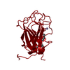
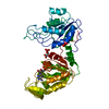
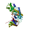
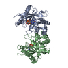
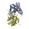
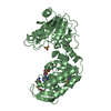
 Archaeoglobus fulgidus (archaea)
 X-RAY DIFFRACTION /
  Authors
 United States, 1items
  Citation
 Structure visualization
 Downloads & links
 Other downloads
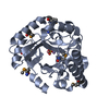


 PDBj
PDBj
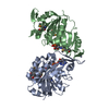
 Assembly
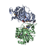


 Mass: 427.201 Da / Num. of mol.: 2 / Source method: obtained synthetically / Formula: C10H15N5O10P2 / Comment: ADP, energy-carrying molecule*YM
 Mass: 58.933 Da / Num. of mol.: 1 / Source method: obtained synthetically / Formula: Co
 Mass: 18.015 Da / Num. of mol.: 8 / Source method: isolated from a natural source / Formula: H2O
 Sample preparation
 Processing