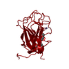
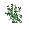
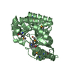
- PDB-6nlr: Crystal structure of the putative histidinol phosphatase hisK fro... -
+
Open data
ID or keywords:
Loading...
-
Basic information
Entry
Database: PDB / ID: 6nlr
Title
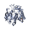
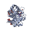
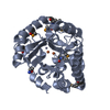
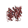
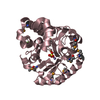
Crystal structure of the putative histidinol phosphatase hisK from Listeria monocytogenes with trinuclear metals determined by PIXE revealing sulphate ion in active site. Based on PIXE analysis and original date from 3DCP
THIS ENTRY 6NLR REFLECTS AN ALTERNATIVE MODELING OF THE ORIGINAL DATA IN 3DCP, DETERMINED BY S.M. ...THIS ENTRY 6NLR REFLECTS AN ALTERNATIVE MODELING OF THE ORIGINAL DATA IN 3DCP, DETERMINED BY S.M.VOROBIEV,M.SU,J.SEETHARAMAN,L.ZHAO,L.MAO,E.L.FOOTE,R.XIAO,R.NAIR,M.C.BARAN,T.B.ACTON,B.ROST,G.T.MONTELIONE,J.F.HUNT,L.TONG,NORTHEAST STRUCTURAL GENOMICS CONSORTIUM (NESG)
Mass: 18.015 Da / Num. of mol.: 529 / Source method: isolated from a natural source / Formula: H2O
-
Details
Has protein modification
Y
-
Experimental details
-
Experiment
Experiment
Method: X-RAY DIFFRACTION / Number of used crystals: 1
-
Sample preparation
Crystal
Density Matthews: 2.58 Å3/Da / Density % sol: 52.23 % / Description: AUTHOR USED THE SF DATA FROM ENTRY 3DCP.
Crystal grow
Temperature: 291 K / Method: vapor diffusion, hanging drop / pH: 6.2 Details: 15% PEG 8000, 0.17 M SODIUM ACETATE, 0.01 M L-CYSTEINE, 0.1 M MES PH 6.2, VAPOR DIFFUSION, HANGING DROP, TEMPERATURE 291K
-
Data collection
Diffraction
Mean temperature: 100 K / Serial crystal experiment: N
Protocol: SINGLE WAVELENGTH / Monochromatic (M) / Laue (L): M / Scattering type: x-ray
Radiation wavelength
Wavelength: 0.97931 Å / Relative weight: 1
Reflection
Resolution: 2.1→41.6 Å / Num. obs: 114574 / % possible obs: 99.8 % / Observed criterion σ(F): 2 / Redundancy: 3.9 % / Rmerge(I) obs: 0.106 / Net I/σ(I): 23.6
Reflection shell
Resolution: 2.1→2.18 Å / Redundancy: 3.8 % / Rmerge(I) obs: 0.272 / Mean I/σ(I) obs: 4.4 / Num. unique obs: 12134 / % possible all: 98.4
-
Processing
Software
Name
Version
Classification
REFMAC
5.8.0241
refinement
HKL-2000
datareduction
HKL-2000
datascaling
SHELXDE
phasing
RESOLVE
phasing
Refinement
Method to determine structure: SAD / Resolution: 2.1→41.6 Å / Cor.coef. Fo:Fc: 0.967 / Cor.coef. Fo:Fc free: 0.956 / SU B: 7.658 / SU ML: 0.107 / Cross valid method: THROUGHOUT / ESU R: 0.173 / ESU R Free: 0.144 / Stereochemistry target values: MAXIMUM LIKELIHOOD / Details: HYDROGENS HAVE BEEN ADDED IN THE RIDING POSITIONS
Rfactor
Num. reflection
% reflection
Selection details
Rfree
0.18583
2868
4.9 %
RANDOM
Rwork
0.15488
-
-
-
obs
0.15638
55392
98.4 %
-
Solvent computation
Ion probe radii: 0.7 Å / Shrinkage radii: 0.7 Å / VDW probe radii: 1 Å / Solvent model: MASK
 Movie
Movie Controller

 Yorodumi
 Open data
 Basic information
 Components
 Keywords
 Function and homology information
 Listeria monocytogenes serotype 4b str. H7858 (bacteria)
 X-RAY DIFFRACTION /
  Authors
 United States, 1items
  Citation
 Structure visualization
 Downloads & links
 Other downloads
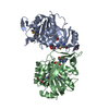


 PDBj
PDBj
 Assembly


 Mass: 54.938 Da / Num. of mol.: 3 / Source method: obtained synthetically / Formula: Mn
 Mass: 58.933 Da / Num. of mol.: 3 / Source method: obtained synthetically / Formula: Co
 Mass: 96.063 Da / Num. of mol.: 3 / Source method: obtained synthetically / Formula: SO4
 Mass: 55.845 Da / Num. of mol.: 3 / Source method: obtained synthetically / Formula: Fe
 Mass: 40.078 Da / Num. of mol.: 2 / Source method: obtained synthetically / Formula: Ca
 Sample preparation
 Processing