 Movie
Movie Controller

[English] 日本語
 Yorodumi
- PDB-6bxs: Crystal structure of Toxoplasma gondii Mitochondrial Association ... -
+ Open data

- Basic information
| Entry | Database: PDB / ID: 6bxs | ||||||
|---|---|---|---|---|---|---|---|
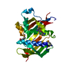
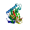
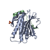
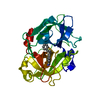
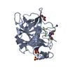
| Title | Crystal structure of Toxoplasma gondii Mitochondrial Association Factor 1 A (MAF1A) | ||||||
 Components Components | mitochondrial association factor 1 | ||||||
 Keywords Keywords | MEMBRANE PROTEIN / macro domain / ADPribose / neofunctionalize | ||||||
| Function / homology | symbiont-containing vacuole membrane / Mitochondrial association factor 1 form a1 Function and homology information Function and homology information | ||||||
| Biological species |  | ||||||
| Method |  X-RAY DIFFRACTION / SYNCHROTRON / MOLECULAR REPLACEMENT / molecular replacement / Resolution: 2.1 Å X-RAY DIFFRACTION / SYNCHROTRON / MOLECULAR REPLACEMENT / molecular replacement / Resolution: 2.1 Å | ||||||
 Authors Authors | Ramaswamy, R. / Boulanger, M.J. | ||||||
| Funding support |  Canada, 1items Canada, 1items
| ||||||
 Citation Citation | Journal: Mol. Microbiol. / Year: 2018 Title: A Toxoplasma gondii locus required for the direct manipulation of host mitochondria has maintained multiple ancestral functions. Authors: Blank, M.L. / Parker, M.L. / Ramaswamy, R. / Powell, C.J. / English, E.D. / Adomako-Ankomah, Y. / Pernas, L.F. / Workman, S.D. / Boothroyd, J.C. / Boulanger, M.J. / Boyle, J.P. | ||||||
| History |
|
- Structure visualization
| Structure viewer | Molecule: MolmilJmol/JSmol |
|---|
- Downloads & links
-Download
| PDBx/mmCIF format | 6bxs.cif.gz | 169.9 KB | Display | PDBx/mmCIF format |
|---|---|---|---|---|
| PDB format | pdb6bxs.ent.gz | 133.8 KB | Display | PDB format |
| PDBx/mmJSON format | 6bxs.json.gz | Tree view | PDBx/mmJSON format | |
| Others |  Other downloads Other downloads |
-Validation report
| Arichive directory | https://data.pdbj.org/pub/pdb/validation_reports/bx/6bxsftp://data.pdbj.org/pub/pdb/validation_reports/bx/6bxs | HTTPS FTP |
|---|
-Related structure data
| Related structure data | 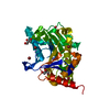 6bxrSC 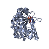 6bxtC 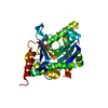 6bxwC S: Starting model for refinement C: citing same article ( |
|---|---|
| Similar structure data |


-Links
 PDBj
PDBj- Assembly
| Deposited unit | 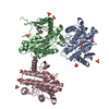
| ||||||||
|---|---|---|---|---|---|---|---|---|---|
| 1 | 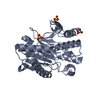
| ||||||||
| 2 | 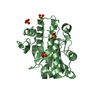
| ||||||||
| 3 | 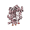
| ||||||||
| Unit cell |
|
-Components
| #1: Protein | Mass: 30857.760 Da / Num. of mol.: 3 Source method: isolated from a genetically manipulated source Source: (gene. exp.)  #2: Chemical | ChemComp-SO4 /   Mass: 96.063 Da / Num. of mol.: 14 / Source method: obtained synthetically / Formula: SO4 Mass: 96.063 Da / Num. of mol.: 14 / Source method: obtained synthetically / Formula: SO4#3: Water | ChemComp-HOH / |  Mass: 18.015 Da / Num. of mol.: 159 / Source method: isolated from a natural source / Formula: H2O Mass: 18.015 Da / Num. of mol.: 159 / Source method: isolated from a natural source / Formula: H2O |
|---|
-Experimental details
-Experiment
| Experiment | Method: X-RAY DIFFRACTION / Number of used crystals: 1 |
|---|
- Sample preparation
| Crystal | Density Matthews: 2.43 Å3/Da / Density % sol: 49.36 % |
|---|---|
| Crystal grow | Temperature: 291 K / Method: vapor diffusion, sitting drop / pH: 7 Details: 0.9 M ammonium sulfate, 0.1M Hepes pH 7.0, 0.5% PEG8000, 3% 2- methyl- 2,4- pentanediol |
-Data collection
| Diffraction | Mean temperature: 100 K |
|---|---|
| Diffraction source | Source: SYNCHROTRON / Site: CLSI / Beamline: 08ID-1 / Wavelength: 0.9794 Å |
| Detector | Type: RAYONIX MX300HE / Detector: CCD / Date: May 27, 2014 Details: Vertical Focusing Mirror: ultra-low expansion (ULE) titanium siliicate flat mirror with Pt, Uncoated, and Pd strips |
| Radiation | Monochromator: ACCEL/BRUKER double crystal monochromator (DCM) Protocol: SINGLE WAVELENGTH / Monochromatic (M) / Laue (L): M / Scattering type: x-ray |
| Radiation wavelength | Wavelength: 0.9794 Å / Relative weight: 1 |
| Reflection | Resolution: 2.1→48.86 Å / Num. obs: 51862 / % possible obs: 98.5 % / Redundancy: 3.8 % / Rmerge(I) obs: 0.08 / Net I/σ(I): 11.5 |
| Reflection shell | Resolution: 2.1→2.16 Å / Redundancy: 3.6 % / Rmerge(I) obs: 0.444 / Mean I/σ(I) obs: 3 / % possible all: 92.4 |
-Phasing
| Phasing | Method: molecular replacement |
|---|
- Processing
| Software |
| ||||||||||||||||||||||||||||||||||||||||||||||||||||||||||||||||||||||||||||||||||||||||||||||||||||||||||||||||||||||||||||||||||||||||||||
|---|---|---|---|---|---|---|---|---|---|---|---|---|---|---|---|---|---|---|---|---|---|---|---|---|---|---|---|---|---|---|---|---|---|---|---|---|---|---|---|---|---|---|---|---|---|---|---|---|---|---|---|---|---|---|---|---|---|---|---|---|---|---|---|---|---|---|---|---|---|---|---|---|---|---|---|---|---|---|---|---|---|---|---|---|---|---|---|---|---|---|---|---|---|---|---|---|---|---|---|---|---|---|---|---|---|---|---|---|---|---|---|---|---|---|---|---|---|---|---|---|---|---|---|---|---|---|---|---|---|---|---|---|---|---|---|---|---|---|---|---|---|
| Refinement | Method to determine structure: MOLECULAR REPLACEMENT Starting model: 6BXR Resolution: 2.1→48.86 Å / SU ML: 0.24 / Cross valid method: FREE R-VALUE / σ(F): 1.35 / Phase error: 25.92
| ||||||||||||||||||||||||||||||||||||||||||||||||||||||||||||||||||||||||||||||||||||||||||||||||||||||||||||||||||||||||||||||||||||||||||||
| Solvent computation | Shrinkage radii: 0.9 Å / VDW probe radii: 1.11 Å | ||||||||||||||||||||||||||||||||||||||||||||||||||||||||||||||||||||||||||||||||||||||||||||||||||||||||||||||||||||||||||||||||||||||||||||
| Displacement parameters | Biso max: 112.37 Å2 / Biso mean: 33.6138 Å2 / Biso min: 15.37 Å2 | ||||||||||||||||||||||||||||||||||||||||||||||||||||||||||||||||||||||||||||||||||||||||||||||||||||||||||||||||||||||||||||||||||||||||||||
| Refinement step | Cycle: final / Resolution: 2.1→48.86 Å
| ||||||||||||||||||||||||||||||||||||||||||||||||||||||||||||||||||||||||||||||||||||||||||||||||||||||||||||||||||||||||||||||||||||||||||||
| Refine LS restraints |
| ||||||||||||||||||||||||||||||||||||||||||||||||||||||||||||||||||||||||||||||||||||||||||||||||||||||||||||||||||||||||||||||||||||||||||||
| LS refinement shell | Refine-ID: X-RAY DIFFRACTION / Total num. of bins used: 19
|
