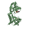
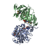
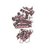
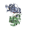
[English] 日本語
Yorodumi
- PDB-5lba: Crystal structure of human RECQL5 helicase in complex with DSPL f... -

+
Open data


ID or keywords:

Loading...

-
Basic information

Entry
Database: PDB / ID: 5lba
TitleCrystal structure of human RECQL5 helicase in complex with DSPL fragment(1-cyclohexyl-3-(oxolan-2-ylmethyl)urea, SGC - Diamond XChem I04-1 fragment screening.
ComponentsATP-dependent DNA helicase Q5
KeywordsHYDROLASE / Helicase / RecQ / Transcription / Fragment screening / Structural Genomics / Structural Genomics Consortium / SGC / DIAMOND I04-1 XCHEM"
Function / homology
Function and homology information


mitotic DNA-templated DNA replication / chromosome separation / cellular response to camptothecin / DNA 3'-5' helicase / four-way junction helicase activity / replication-born double-strand break repair via sister chromatid exchange / 3'-5' DNA helicase activity / transcription preinitiation complex / DNA metabolic process / negative regulation of double-strand break repair via homologous recombination ...mitotic DNA-templated DNA replication / chromosome separation / cellular response to camptothecin / DNA 3'-5' helicase / four-way junction helicase activity / replication-born double-strand break repair via sister chromatid exchange / 3'-5' DNA helicase activity / transcription preinitiation complex / DNA metabolic process / negative regulation of double-strand break repair via homologous recombination / DNA unwinding involved in DNA replication / RNA polymerase II complex binding / negative regulation of transcription elongation by RNA polymerase II / DNA helicase activity / replication fork / helicase activity / double-strand break repair via homologous recombination / cellular response to xenobiotic stimulus / mitotic cell cycle / chromosome / DNA replication / nucleic acid binding / cell division / DNA repair / ATP hydrolysis activity / nucleoplasm / ATP binding / identical protein binding / nucleus / cytosol / cytoplasm
Similarity search - Function
RecQ helicase-like 5 / RecQ helicase protein-like 5 (RecQ5) / ATP-dependent DNA helicase RecQ, zinc-binding domain / RecQ zinc-binding / DNA helicase, ATP-dependent, RecQ type / DNA/RNA helicase, ATP-dependent, DEAH-box type, conserved site / DEAH-box subfamily ATP-dependent helicases signature. / DEAD/DEAH box helicase / DEAD/DEAH box helicase domain / Helicase conserved C-terminal domain ...RecQ helicase-like 5 / RecQ helicase protein-like 5 (RecQ5) / ATP-dependent DNA helicase RecQ, zinc-binding domain / RecQ zinc-binding / DNA helicase, ATP-dependent, RecQ type / DNA/RNA helicase, ATP-dependent, DEAH-box type, conserved site / DEAH-box subfamily ATP-dependent helicases signature. / DEAD/DEAH box helicase / DEAD/DEAH box helicase domain / Helicase conserved C-terminal domain / helicase superfamily c-terminal domain / Superfamilies 1 and 2 helicase C-terminal domain profile. / Superfamilies 1 and 2 helicase ATP-binding type-1 domain profile. / DEAD-like helicases superfamily / Helicase, C-terminal / Helicase superfamily 1/2, ATP-binding domain / P-loop containing nucleotide triphosphate hydrolases / P-loop containing nucleoside triphosphate hydrolase / Rossmann fold / 3-Layer(aba) Sandwich / Alpha Beta
Similarity search - Domain/homology
1-cyclohexyl-3-[[(2~{R})-oxolan-2-yl]methyl]urea / ADENOSINE-5'-DIPHOSPHATE / ATP-dependent DNA helicase Q5
Similarity search - Component
Biological speciesHomo sapiens (human)
MethodX-RAY DIFFRACTION / SYNCHROTRON / FOURIER SYNTHESIS / Resolution: 2.5 Å
AuthorsNewman, J.A. / Aitkenhead, H. / Talon, R. / Savitsky, P. / Krojer, T. / von Delft, F. / Arrowsmith, C.H. / Edwards, A.M. / Bountra, C. / Gileadi, O. / Structural Genomics Consortium (SGC)
Funding support United Kingdom, 1items
OrganizationGrant numberCountry
Wellcome Trust United Kingdom
CitationJournal: To be published
Title: Crystal structure of human RECQL5 helicase in complex with 3D fragment (1-cyclohexyl-3-(oxolan-2-ylmethyl)urea)
Authors: Newman, J.A. / Talon, R. / Aitkenhead, H. / Savitsky, P. / Krojer, T. / von Delft, F. / Arrowsmith, C.H. / Edwards, A.M. / Bountra, C. / Gileadi, O.
History
DepositionJun 15, 2016Deposition site: PDBE / Processing site: PDBE
Revision 1.0Jul 6, 2016Provider: repository / Type: Initial release
Revision 1.1Jan 24, 2018Group: Database references / Structure summary / Category: audit_author / citation_author / Item: _audit_author.name / _citation_author.name
Revision 1.2Jan 10, 2024Group: Data collection / Database references ...Data collection / Database references / Derived calculations / Refinement description
Category: chem_comp_atom / chem_comp_bond ...chem_comp_atom / chem_comp_bond / database_2 / pdbx_initial_refinement_model / pdbx_struct_conn_angle / struct_conn
Item: _database_2.pdbx_DOI / _database_2.pdbx_database_accession ..._database_2.pdbx_DOI / _database_2.pdbx_database_accession / _pdbx_struct_conn_angle.ptnr1_auth_asym_id / _pdbx_struct_conn_angle.ptnr1_auth_comp_id / _pdbx_struct_conn_angle.ptnr1_auth_seq_id / _pdbx_struct_conn_angle.ptnr1_label_asym_id / _pdbx_struct_conn_angle.ptnr1_label_atom_id / _pdbx_struct_conn_angle.ptnr1_label_comp_id / _pdbx_struct_conn_angle.ptnr1_label_seq_id / _pdbx_struct_conn_angle.ptnr2_auth_asym_id / _pdbx_struct_conn_angle.ptnr2_auth_comp_id / _pdbx_struct_conn_angle.ptnr2_auth_seq_id / _pdbx_struct_conn_angle.ptnr2_label_asym_id / _pdbx_struct_conn_angle.ptnr2_label_atom_id / _pdbx_struct_conn_angle.ptnr2_label_comp_id / _pdbx_struct_conn_angle.ptnr3_auth_asym_id / _pdbx_struct_conn_angle.ptnr3_auth_comp_id / _pdbx_struct_conn_angle.ptnr3_auth_seq_id / _pdbx_struct_conn_angle.ptnr3_label_asym_id / _pdbx_struct_conn_angle.ptnr3_label_atom_id / _pdbx_struct_conn_angle.ptnr3_label_comp_id / _pdbx_struct_conn_angle.ptnr3_label_seq_id / _pdbx_struct_conn_angle.value / _struct_conn.pdbx_dist_value / _struct_conn.ptnr1_auth_asym_id / _struct_conn.ptnr1_auth_comp_id / _struct_conn.ptnr1_auth_seq_id / _struct_conn.ptnr1_label_asym_id / _struct_conn.ptnr1_label_atom_id / _struct_conn.ptnr1_label_comp_id / _struct_conn.ptnr1_label_seq_id / _struct_conn.ptnr2_auth_asym_id / _struct_conn.ptnr2_auth_comp_id / _struct_conn.ptnr2_auth_seq_id / _struct_conn.ptnr2_label_asym_id / _struct_conn.ptnr2_label_atom_id / _struct_conn.ptnr2_label_comp_id

-
Structure visualization

Structure viewerMolecule:
MolmilJmol/JSmol

Downloads & links

-
Assembly

Deposited unit
B: ATP-dependent DNA helicase Q5
A: ATP-dependent DNA helicase Q5
C: ATP-dependent DNA helicase Q5
D: ATP-dependent DNA helicase Q5
hetero molecules


Theoretical massNumber of molelcules
Total (without water)200,67720
Polymers198,1494
Non-polymers2,52816
Water6,089338
1
B: ATP-dependent DNA helicase Q5
hetero molecules


Theoretical massNumber of molelcules
Total (without water)50,3596
Polymers49,5371
Non-polymers8215
Water181
TypeNameSymmetry operationNumber
identity operation1_555x,y,z1
2
A: ATP-dependent DNA helicase Q5
hetero molecules


Theoretical massNumber of molelcules
Total (without water)50,1325
Polymers49,5371
Non-polymers5954
Water181
TypeNameSymmetry operationNumber
identity operation1_555x,y,z1
3
C: ATP-dependent DNA helicase Q5
hetero molecules


Theoretical massNumber of molelcules
Total (without water)50,0544
Polymers49,5371
Non-polymers5173
Water181
TypeNameSymmetry operationNumber
identity operation1_555x,y,z1
4
D: ATP-dependent DNA helicase Q5
hetero molecules


Theoretical massNumber of molelcules
Total (without water)50,1325
Polymers49,5371
Non-polymers5954
Water181
TypeNameSymmetry operationNumber
identity operation1_555x,y,z1
Unit cell
Length a, b, c (Å)63.926, 85.261, 105.915
Angle α, β, γ (deg.)109.90, 90.02, 97.01
Int Tables number1
Space group name H-MP1

-
Components

-
Protein , 1 types, 4 molecules BACD

#1: Protein
ATP-dependent DNA helicase Q5 / DNA helicase / RecQ-like type 5 / RecQ5 / RecQ protein-like 5


Mass: 49537.180 Da / Num. of mol.: 4
Source method: isolated from a genetically manipulated source
Source: (gene. exp.) Homo sapiens (human) / Gene: RECQL5, RECQ5 / Production host: Escherichia coli BL21 (bacteria) / References: UniProt: O94762, DNA helicase

-
Non-polymers , 6 types, 354 molecules

#2: Chemical
ChemComp-MG / MAGNESIUM ION


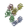
Mass: 24.305 Da / Num. of mol.: 4 / Source method: obtained synthetically / Formula: Mg
#3: Chemical
ChemComp-ZN / ZINC ION


Mass: 65.409 Da / Num. of mol.: 4 / Source method: obtained synthetically / Formula: Zn
#4: Chemical
ChemComp-ADP / ADENOSINE-5'-DIPHOSPHATE / Adenosine diphosphate


Mass: 427.201 Da / Num. of mol.: 4 / Source method: obtained synthetically / Formula: C10H15N5O10P2 / Comment: ADP, energy-carrying molecule*YM
#5: Chemical ChemComp-DMS / DIMETHYL SULFOXIDE / Dimethyl sulfoxide


Mass: 78.133 Da / Num. of mol.: 3 / Source method: obtained synthetically / Formula: C2H6OS / Comment: DMSO, precipitant*YM
#6: Chemical ChemComp-6SV / 1-cyclohexyl-3-[[(2~{R})-oxolan-2-yl]methyl]urea


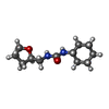
Mass: 226.315 Da / Num. of mol.: 1 / Source method: obtained synthetically / Formula: C12H22N2O2
#7: Water ChemComp-HOH / water / Water


Mass: 18.015 Da / Num. of mol.: 338 / Source method: isolated from a natural source / Formula: H2O

-
Experimental details

-
Experiment

ExperimentMethod: X-RAY DIFFRACTION / Number of used crystals: 1

-
Sample preparation

CrystalDensity Matthews: 2.72 Å3/Da / Density % sol: 54.72 %
Crystal growTemperature: 293 K / Method: vapor diffusion, sitting drop
Details: 0.1M tris pH 9.0 -- 29% PEG3350 -- 0.1M sodium chloride

-
Data collection

DiffractionMean temperature: 100 K
Diffraction sourceSource: SYNCHROTRON / Site: Diamond / Beamline: I04-1 / Wavelength: 0.9282 Å
DetectorType: DECTRIS PILATUS3 6M / Detector: PIXEL / Date: Jan 19, 2016
RadiationProtocol: SINGLE WAVELENGTH / Monochromatic (M) / Laue (L): M / Scattering type: x-ray
Radiation wavelengthWavelength: 0.9282 Å / Relative weight: 1
ReflectionResolution: 2.47→79.49 Å / Num. obs: 69069 / % possible obs: 92.3 % / Redundancy: 2.5 % / Rmerge(I) obs: 0.097 / Net I/σ(I): 5.8
Reflection shellResolution: 2.47→2.53 Å / Redundancy: 2.5 % / Rmerge(I) obs: 0.513 / Mean I/σ(I) obs: 1.3 / % possible all: 94.3

-
Processing

Software
NameVersionClassification
REFMAC5.8.0135refinement
XDSdata reduction
XDSdata scaling
REFMACphasing
RefinementMethod to determine structure: FOURIER SYNTHESIS
Starting model: 5LB5
Resolution: 2.5→79.49 Å / Cor.coef. Fo:Fc: 0.92 / Cor.coef. Fo:Fc free: 0.863 / SU B: 15.593 / SU ML: 0.329 / Cross valid method: THROUGHOUT / ESU R: 0.829 / ESU R Free: 0.37 / Details: HYDROGENS HAVE BEEN ADDED IN THE RIDING POSITIONS
RfactorNum. reflection% reflectionSelection details
Rfree0.30292 3363 5 %RANDOM
Rwork0.23665 ---
obs0.24 63558 92.28 %-
Solvent computationIon probe radii: 0.8 Å / Shrinkage radii: 0.8 Å / VDW probe radii: 1.2 Å
Displacement parametersBiso mean: 48.293 Å2
Baniso -1Baniso -2Baniso -3
1--0.92 Å2-1.61 Å2-0.7 Å2
2--1.47 Å21.54 Å2
3----1.02 Å2
Refinement stepCycle: 1 / Resolution: 2.5→79.49 Å
ProteinNucleic acidLigandSolventTotal
Num. atoms13429 0 144 338 13911
Refine LS restraints
Refine-IDTypeDev idealDev ideal targetNumber
X-RAY DIFFRACTIONr_bond_refined_d0.0120.01913879
X-RAY DIFFRACTIONr_bond_other_d0.0020.0213357
X-RAY DIFFRACTIONr_angle_refined_deg1.4961.97418798
X-RAY DIFFRACTIONr_angle_other_deg0.958330661
X-RAY DIFFRACTIONr_dihedral_angle_1_deg7.67551732
X-RAY DIFFRACTIONr_dihedral_angle_2_deg30.66823.057592
X-RAY DIFFRACTIONr_dihedral_angle_3_deg14.231152366
X-RAY DIFFRACTIONr_dihedral_angle_4_deg14.26215115
X-RAY DIFFRACTIONr_chiral_restr0.0740.22122
X-RAY DIFFRACTIONr_gen_planes_refined0.0070.02115588
X-RAY DIFFRACTIONr_gen_planes_other0.0010.023215
X-RAY DIFFRACTIONr_nbd_refined
X-RAY DIFFRACTIONr_nbd_other
X-RAY DIFFRACTIONr_nbtor_refined
X-RAY DIFFRACTIONr_nbtor_other
X-RAY DIFFRACTIONr_xyhbond_nbd_refined
X-RAY DIFFRACTIONr_xyhbond_nbd_other
X-RAY DIFFRACTIONr_metal_ion_refined
X-RAY DIFFRACTIONr_metal_ion_other
X-RAY DIFFRACTIONr_symmetry_vdw_refined
X-RAY DIFFRACTIONr_symmetry_vdw_other
X-RAY DIFFRACTIONr_symmetry_hbond_refined
X-RAY DIFFRACTIONr_symmetry_hbond_other
X-RAY DIFFRACTIONr_symmetry_metal_ion_refined
X-RAY DIFFRACTIONr_symmetry_metal_ion_other
X-RAY DIFFRACTIONr_mcbond_it3.0624.7276946
X-RAY DIFFRACTIONr_mcbond_other3.0624.7276945
X-RAY DIFFRACTIONr_mcangle_it4.9427.0778672
X-RAY DIFFRACTIONr_mcangle_other4.9417.0778673
X-RAY DIFFRACTIONr_scbond_it2.9064.9986933
X-RAY DIFFRACTIONr_scbond_other2.9064.9986934
X-RAY DIFFRACTIONr_scangle_it
X-RAY DIFFRACTIONr_scangle_other4.8317.37110127
X-RAY DIFFRACTIONr_long_range_B_refined7.6836.61515044
X-RAY DIFFRACTIONr_long_range_B_other7.6836.61815045
X-RAY DIFFRACTIONr_rigid_bond_restr
X-RAY DIFFRACTIONr_sphericity_free
X-RAY DIFFRACTIONr_sphericity_bonded
LS refinement shellResolution: 2.496→2.561 Å / Total num. of bins used: 20
RfactorNum. reflection% reflection
Rfree0.396 264 -
Rwork0.33 4818 -
obs--93.95 %

+
About Yorodumi

-
News

-
Feb 9, 2022. New format data for meta-information of EMDB entries

New format data for meta-information of EMDB entries

  • Version 3 of the EMDB header file is now the official format.
  • The previous official version 1.9 will be removed from the archive.

Related info.:EMDB header

External links:wwPDB to switch to version 3 of the EMDB data model

-
Aug 12, 2020. Covid-19 info

Covid-19 info

URL: https://pdbj.org/emnavi/covid19.php

New page: Covid-19 featured information page in EM Navigator.

Related info.:Covid-19 info / Mar 5, 2020. Novel coronavirus structure data

+
Mar 5, 2020. Novel coronavirus structure data

Novel coronavirus structure data

Related info.:Yorodumi Speices / Aug 12, 2020. Covid-19 info

External links:COVID-19 featured content - PDBj / Molecule of the Month (242):Coronavirus Proteases

+
Jan 31, 2019. EMDB accession codes are about to change! (news from PDBe EMDB page)

EMDB accession codes are about to change! (news from PDBe EMDB page)

  • The allocation of 4 digits for EMDB accession codes will soon come to an end. Whilst these codes will remain in use, new EMDB accession codes will include an additional digit and will expand incrementally as the available range of codes is exhausted. The current 4-digit format prefixed with “EMD-” (i.e. EMD-XXXX) will advance to a 5-digit format (i.e. EMD-XXXXX), and so on. It is currently estimated that the 4-digit codes will be depleted around Spring 2019, at which point the 5-digit format will come into force.
  • The EM Navigator/Yorodumi systems omit the EMD- prefix.

Related info.:Q: What is EMD? / ID/Accession-code notation in Yorodumi/EM Navigator

External links:EMDB Accession Codes are Changing Soon! / Contact to PDBj

+
Jul 12, 2017. Major update of PDB

Major update of PDB

  • wwPDB released updated PDB data conforming to the new PDBx/mmCIF dictionary.
  • This is a major update changing the version number from 4 to 5, and with Remediation, in which all the entries are updated.
  • In this update, many items about electron microscopy experimental information are reorganized (e.g. em_software).
  • Now, EM Navigator and Yorodumi are based on the updated data.

External links:wwPDB Remediation / Enriched Model Files Conforming to OneDep Data Standards Now Available in the PDB FTP Archive

-
Yorodumi

Thousand views of thousand structures

  • Yorodumi is a browser for structure data from EMDB, PDB, SASBDB, etc.
  • This page is also the successor to EM Navigator detail page, and also detail information page/front-end page for Omokage search.
  • The word "yorodu" (or yorozu) is an old Japanese word meaning "ten thousand". "mi" (miru) is to see.

Related info.:EMDB / PDB / SASBDB / Comparison of 3 databanks / Yorodumi Search / Aug 31, 2016. New EM Navigator & Yorodumi / Yorodumi Papers / Jmol/JSmol / Function and homology information / Changes in new EM Navigator and Yorodumi

Read more