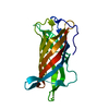
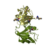
[English] 日本語
Yorodumi
- PDB-6n5w: Crystal structure of the Ca2+/CaM complex with independent peptid... -

+
Open data


ID or keywords:

Loading...

-
Basic information

Entry
Database: PDB / ID: 6n5w
TitleCrystal structure of the Ca2+/CaM complex with independent peptides of Kv7.4 (KCNQ4) A & B domains
Components
  • (Potassium voltage-gated channel subfamily KQT member 4) x 2
  • Calmodulin-1
KeywordsMETAL BINDING PROTEIN / ion channel / calmodulin / KCNQ4 peptides / complex
Function / homology
Function and homology information


Voltage gated Potassium channels / Sensory processing of sound by outer hair cells of the cochlea / CaM pathway / Cam-PDE 1 activation / Sodium/Calcium exchangers / Calmodulin induced events / Sensory processing of sound by inner hair cells of the cochlea / positive regulation of ryanodine-sensitive calcium-release channel activity / Reduction of cytosolic Ca++ levels / Activation of Ca-permeable Kainate Receptor ...Voltage gated Potassium channels / Sensory processing of sound by outer hair cells of the cochlea / CaM pathway / Cam-PDE 1 activation / Sodium/Calcium exchangers / Calmodulin induced events / Sensory processing of sound by inner hair cells of the cochlea / positive regulation of ryanodine-sensitive calcium-release channel activity / Reduction of cytosolic Ca++ levels / Activation of Ca-permeable Kainate Receptor / CREB1 phosphorylation through the activation of CaMKII/CaMKK/CaMKIV cascasde / Loss of phosphorylation of MECP2 at T308 / inner ear morphogenesis / CREB1 phosphorylation through the activation of Adenylate Cyclase / CaMK IV-mediated phosphorylation of CREB / PKA activation / negative regulation of high voltage-gated calcium channel activity / Glycogen breakdown (glycogenolysis) / CLEC7A (Dectin-1) induces NFAT activation / Activation of RAC1 downstream of NMDARs / organelle localization by membrane tethering / negative regulation of ryanodine-sensitive calcium-release channel activity / mitochondrion-endoplasmic reticulum membrane tethering / autophagosome membrane docking / negative regulation of calcium ion export across plasma membrane / regulation of cardiac muscle cell action potential / presynaptic endocytosis / Synthesis of IP3 and IP4 in the cytosol / regulation of cell communication by electrical coupling involved in cardiac conduction / Phase 0 - rapid depolarisation / calcineurin-mediated signaling / Negative regulation of NMDA receptor-mediated neuronal transmission / Unblocking of NMDA receptors, glutamate binding and activation / RHO GTPases activate PAKs / Ion transport by P-type ATPases / Uptake and function of anthrax toxins / regulation of ryanodine-sensitive calcium-release channel activity / Long-term potentiation / protein phosphatase activator activity / Calcineurin activates NFAT / Regulation of MECP2 expression and activity / voltage-gated potassium channel activity / DARPP-32 events / catalytic complex / Smooth Muscle Contraction / detection of calcium ion / potassium channel activity / regulation of cardiac muscle contraction / RHO GTPases activate IQGAPs / regulation of cardiac muscle contraction by regulation of the release of sequestered calcium ion / calcium channel inhibitor activity / cellular response to interferon-beta / Protein methylation / presynaptic cytosol / Activation of AMPK downstream of NMDARs / Ion homeostasis / regulation of release of sequestered calcium ion into cytosol by sarcoplasmic reticulum / eNOS activation / titin binding / Tetrahydrobiopterin (BH4) synthesis, recycling, salvage and regulation / sperm midpiece / voltage-gated potassium channel complex / regulation of calcium-mediated signaling / potassium ion transmembrane transport / calcium channel complex / substantia nigra development / FCERI mediated Ca+2 mobilization / Ras activation upon Ca2+ influx through NMDA receptor / regulation of heart rate / FCGR3A-mediated IL10 synthesis / basal plasma membrane / calyx of Held / Antigen activates B Cell Receptor (BCR) leading to generation of second messengers / adenylate cyclase activator activity / sarcomere / VEGFR2 mediated cell proliferation / regulation of cytokinesis / protein serine/threonine kinase activator activity / VEGFR2 mediated vascular permeability / Translocation of SLC2A4 (GLUT4) to the plasma membrane / spindle microtubule / positive regulation of receptor signaling pathway via JAK-STAT / sensory perception of sound / RAF activation / Transcriptional activation of mitochondrial biogenesis / potassium ion transport / Stimuli-sensing channels / cellular response to type II interferon / long-term synaptic potentiation / response to calcium ion / RAS processing / spindle pole / Signaling by RAF1 mutants / Signaling by moderate kinase activity BRAF mutants / Paradoxical activation of RAF signaling by kinase inactive BRAF / Signaling downstream of RAS mutants / calcium-dependent protein binding / G2/M transition of mitotic cell cycle / Signaling by BRAF and RAF1 fusions / Inactivation, recovery and regulation of the phototransduction cascade
Similarity search - Function
Potassium channel, voltage dependent, KCNQ / Potassium channel, voltage dependent, KCNQ, C-terminal / KCNQ voltage-gated potassium channel / EF-hand / : / Recoverin; domain 1 / EF-hand domain pair / EF-hand, calcium binding motif / EF-Hand 1, calcium-binding site / EF-hand calcium-binding domain. ...Potassium channel, voltage dependent, KCNQ / Potassium channel, voltage dependent, KCNQ, C-terminal / KCNQ voltage-gated potassium channel / EF-hand / : / Recoverin; domain 1 / EF-hand domain pair / EF-hand, calcium binding motif / EF-Hand 1, calcium-binding site / EF-hand calcium-binding domain. / EF-hand calcium-binding domain profile. / EF-hand domain / Ion transport domain / Ion transport protein / EF-hand domain pair / Orthogonal Bundle / Mainly Alpha
Similarity search - Domain/homology
Calmodulin-1 / Potassium voltage-gated channel subfamily KQT member 4
Similarity search - Component
Biological speciesHomo sapiens (human)
MethodX-RAY DIFFRACTION / SYNCHROTRON / MOLECULAR REPLACEMENT / Resolution: 2.15 Å
AuthorsTaylor, A.B. / Archer, C.R. / Shapiro, M.S.
Funding support United States, 3items
OrganizationGrant numberCountry
National Institutes of Health/National Institute of Neurological Disorders and Stroke (NIH/NINDS)R01 NS043394 United States
National Institutes of Health/National Institute of Neurological Disorders and Stroke (NIH/NINDS)R01 NS094461 United States
National Institutes of Health/National Heart, Lung, and Blood Institute (NIH/NHLBI)T32 HL007446 United States
CitationJournal: J. Biol. Chem. / Year: 2019
Title: A mutually induced conformational fit underlies Ca2+-directed interactions between calmodulin and the proximal C terminus of KCNQ4 K+channels.
Authors: Archer, C.R. / Enslow, B.T. / Taylor, A.B. / De la Rosa, V. / Bhattacharya, A. / Shapiro, M.S.
History
DepositionNov 22, 2018Deposition site: RCSB / Processing site: RCSB
Revision 1.0Mar 13, 2019Provider: repository / Type: Initial release
Revision 1.1Apr 24, 2019Group: Data collection / Database references / Category: citation
Item: _citation.journal_volume / _citation.page_first ..._citation.journal_volume / _citation.page_first / _citation.page_last / _citation.title
Revision 1.2Dec 4, 2019Group: Author supporting evidence / Category: pdbx_audit_support / Item: _pdbx_audit_support.funding_organization
Revision 1.3Oct 11, 2023Group: Data collection / Database references / Refinement description
Category: chem_comp_atom / chem_comp_bond ...chem_comp_atom / chem_comp_bond / database_2 / pdbx_initial_refinement_model
Item: _database_2.pdbx_DOI / _database_2.pdbx_database_accession

-
Structure visualization

Structure viewerMolecule:
MolmilJmol/JSmol

Downloads & links

-
Assembly

Deposited unit
A: Potassium voltage-gated channel subfamily KQT member 4
B: Potassium voltage-gated channel subfamily KQT member 4
C: Calmodulin-1
hetero molecules


Theoretical massNumber of molelcules
Total (without water)23,2165
Polymers23,1363
Non-polymers802
Water41423
1


  • Idetical with deposited unit
  • defined by author&software
TypeNameSymmetry operationNumber
identity operation1_555x,y,z1
Buried area4990 Å2
ΔGint-72 kcal/mol
Surface area10080 Å2
MethodPISA
Unit cell
Length a, b, c (Å)43.510, 130.570, 36.160
Angle α, β, γ (deg.)90.00, 90.00, 90.00
Int Tables number18
Space group name H-MP21212

-
Components

#1: Protein/peptide Potassium voltage-gated channel subfamily KQT member 4 / KQT-like 4 / Potassium channel subunit alpha KvLQT4 / Voltage-gated potassium channel subunit Kv7.4


Mass: 3217.747 Da / Num. of mol.: 1 / Source method: obtained synthetically / Source: (synth.) Homo sapiens (human) / References: UniProt: P56696
#2: Protein/peptide Potassium voltage-gated channel subfamily KQT member 4 / KQT-like 4 / Potassium channel subunit alpha KvLQT4 / Voltage-gated potassium channel subunit Kv7.4


Mass: 3065.826 Da / Num. of mol.: 1 / Source method: obtained synthetically / Source: (synth.) Homo sapiens (human) / References: UniProt: P56696
#3: Protein Calmodulin-1


Mass: 16852.545 Da / Num. of mol.: 1
Source method: isolated from a genetically manipulated source
Source: (gene. exp.) Homo sapiens (human) / Gene: CALM1, CALM, CAM, CAM1 / Plasmid: pETGQ.HCaM / Production host: Escherichia coli BL21 (bacteria) / References: UniProt: P0DP23
#4: Chemical ChemComp-CA / CALCIUM ION


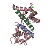
Mass: 40.078 Da / Num. of mol.: 2 / Source method: obtained synthetically / Formula: Ca
#5: Water ChemComp-HOH / water


Mass: 18.015 Da / Num. of mol.: 23 / Source method: isolated from a natural source / Formula: H2O

-
Experimental details

-
Experiment

ExperimentMethod: X-RAY DIFFRACTION / Number of used crystals: 1

-
Sample preparation

CrystalDensity Matthews: 2.22 Å3/Da / Density % sol: 44.59 %
Crystal growTemperature: 295 K / Method: vapor diffusion, sitting drop / Details: 1.3 M sodium citrate, 0.1 M HEPES pH 7.0

-
Data collection

DiffractionMean temperature: 100 K / Serial crystal experiment: N
Diffraction sourceSource: SYNCHROTRON / Site: APS / Beamline: 24-ID-E / Wavelength: 0.97918 Å
DetectorType: ADSC QUANTUM 315 / Detector: CCD / Date: Mar 26, 2016
RadiationProtocol: SINGLE WAVELENGTH / Monochromatic (M) / Laue (L): M / Scattering type: x-ray
Radiation wavelengthWavelength: 0.97918 Å / Relative weight: 1
ReflectionResolution: 2.15→43.52 Å / Num. obs: 11851 / % possible obs: 100 % / Redundancy: 7.9 % / Biso Wilson estimate: 45.6 Å2 / CC1/2: 0.995 / Rpim(I) all: 0.06 / Rsym value: 0.157 / Net I/σ(I): 8.1
Reflection shellResolution: 2.15→2.27 Å / Redundancy: 7.9 % / Mean I/σ(I) obs: 2 / Num. unique obs: 1702 / CC1/2: 0.535 / Rpim(I) all: 0.404 / Rsym value: 1.073 / % possible all: 100

-
Processing

Software
NameVersionClassification
PHENIX(1.13_2998: ???)refinement
XDSdata reduction
XDSdata scaling
PHASERphasing
RefinementMethod to determine structure: MOLECULAR REPLACEMENT
Starting model: 4UMO
Resolution: 2.15→41.278 Å / SU ML: 0.34 / Cross valid method: THROUGHOUT / σ(F): 1.36 / Phase error: 30.3
RfactorNum. reflection% reflection
Rfree0.2739 1179 9.98 %
Rwork0.2325 --
obs0.2367 11810 99.89 %
Solvent computationShrinkage radii: 0.9 Å / VDW probe radii: 1.11 Å
Displacement parametersBiso mean: 59.9 Å2
Refinement stepCycle: LAST / Resolution: 2.15→41.278 Å
ProteinNucleic acidLigandSolventTotal
Num. atoms1497 0 2 23 1522
Refine LS restraints
Refine-IDTypeDev idealNumber
X-RAY DIFFRACTIONf_bond_d0.011514
X-RAY DIFFRACTIONf_angle_d1.1272032
X-RAY DIFFRACTIONf_dihedral_angle_d23.281581
X-RAY DIFFRACTIONf_chiral_restr0.077227
X-RAY DIFFRACTIONf_plane_restr0.005269
LS refinement shell
Resolution (Å)Rfactor RfreeNum. reflection RfreeRfactor RworkNum. reflection RworkRefine-ID% reflection obs (%)
2.15-2.24790.39211180.34251326X-RAY DIFFRACTION100
2.2479-2.36640.34331770.31811259X-RAY DIFFRACTION100
2.3664-2.51460.29851180.2991319X-RAY DIFFRACTION100
2.5146-2.70870.35131770.27961284X-RAY DIFFRACTION100
2.7087-2.98120.34111180.2711360X-RAY DIFFRACTION100
2.9812-3.41240.32741770.26121281X-RAY DIFFRACTION100
3.4124-4.29860.26941180.19431386X-RAY DIFFRACTION100
4.2986-41.28610.20351760.19651416X-RAY DIFFRACTION99
Refinement TLS params.

Method: refined / Refine-ID: X-RAY DIFFRACTION

IDL112)L122)L132)L222)L232)L332)S11 (Å °)S12 (Å °)S13 (Å °)S21 (Å °)S22 (Å °)S23 (Å °)S31 (Å °)S32 (Å °)S33 (Å °)T112)T122)T132)T222)T232)T332)Origin x (Å)Origin y (Å)Origin z (Å)
11.01060.8776-0.43191.3045-0.48320.4599-0.26580.0964-0.1637-0.39480.6325-0.0231-0.3436-0.56460.13660.50730.1133-0.01370.3210.09960.2434-18.605120.8231-5.8396
20.2397-0.1532-0.20430.15020.22420.33990.10140.05030.00950.5764-0.3538-0.4282-0.1181-0.054200.3744-0.03520.01230.37470.02930.305-14.042310.4558-9.6824
31.7814-0.1787-0.57661.02010.40420.7380.06310.2590.0710.1034-0.0651-0.2743-0.08820.003-00.2917-0.02690.03840.34910.05950.3907-9.14946.1267-15.8142
41.6641-0.2153-0.57710.58470.92964.57170.4137-0.18140.14460.2120.27250.1581-1.3472-0.25062.06270.66730.13210.16250.4170.23920.5055-19.698525.87061.7816
Refinement TLS group
IDRefine-IDRefine TLS-IDSelection details
1X-RAY DIFFRACTION1chain 'A' and (resid 338 through 356 )
2X-RAY DIFFRACTION2chain 'B' and (resid 525 through 549 )
3X-RAY DIFFRACTION3chain 'C' and (resid 4 through 78 )
4X-RAY DIFFRACTION4chain 'C' and (resid 79 through 147 )

+
About Yorodumi

-
News

-
Feb 9, 2022. New format data for meta-information of EMDB entries

New format data for meta-information of EMDB entries

  • Version 3 of the EMDB header file is now the official format.
  • The previous official version 1.9 will be removed from the archive.

Related info.:EMDB header

External links:wwPDB to switch to version 3 of the EMDB data model

-
Aug 12, 2020. Covid-19 info

Covid-19 info

URL: https://pdbj.org/emnavi/covid19.php

New page: Covid-19 featured information page in EM Navigator.

Related info.:Covid-19 info / Mar 5, 2020. Novel coronavirus structure data

+
Mar 5, 2020. Novel coronavirus structure data

Novel coronavirus structure data

Related info.:Yorodumi Speices / Aug 12, 2020. Covid-19 info

External links:COVID-19 featured content - PDBj / Molecule of the Month (242):Coronavirus Proteases

+
Jan 31, 2019. EMDB accession codes are about to change! (news from PDBe EMDB page)

EMDB accession codes are about to change! (news from PDBe EMDB page)

  • The allocation of 4 digits for EMDB accession codes will soon come to an end. Whilst these codes will remain in use, new EMDB accession codes will include an additional digit and will expand incrementally as the available range of codes is exhausted. The current 4-digit format prefixed with “EMD-” (i.e. EMD-XXXX) will advance to a 5-digit format (i.e. EMD-XXXXX), and so on. It is currently estimated that the 4-digit codes will be depleted around Spring 2019, at which point the 5-digit format will come into force.
  • The EM Navigator/Yorodumi systems omit the EMD- prefix.

Related info.:Q: What is EMD? / ID/Accession-code notation in Yorodumi/EM Navigator

External links:EMDB Accession Codes are Changing Soon! / Contact to PDBj

+
Jul 12, 2017. Major update of PDB

Major update of PDB

  • wwPDB released updated PDB data conforming to the new PDBx/mmCIF dictionary.
  • This is a major update changing the version number from 4 to 5, and with Remediation, in which all the entries are updated.
  • In this update, many items about electron microscopy experimental information are reorganized (e.g. em_software).
  • Now, EM Navigator and Yorodumi are based on the updated data.

External links:wwPDB Remediation / Enriched Model Files Conforming to OneDep Data Standards Now Available in the PDB FTP Archive

-
Yorodumi

Thousand views of thousand structures

  • Yorodumi is a browser for structure data from EMDB, PDB, SASBDB, etc.
  • This page is also the successor to EM Navigator detail page, and also detail information page/front-end page for Omokage search.
  • The word "yorodu" (or yorozu) is an old Japanese word meaning "ten thousand". "mi" (miru) is to see.

Related info.:EMDB / PDB / SASBDB / Comparison of 3 databanks / Yorodumi Search / Aug 31, 2016. New EM Navigator & Yorodumi / Yorodumi Papers / Jmol/JSmol / Function and homology information / Changes in new EM Navigator and Yorodumi

Read more