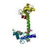
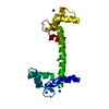
Entry Database : PDB / ID : 6mueTitle Voltage-gated sodium channel NaV1.4 IQ domain in complex with Ca2+/Calmodulin Calmodulin-1 Sodium channel protein type 4 subunit alpha Keywords / / / / Function / homology Function Domain/homology Component
/ / / / / / / / / / / / / / / / / / / / / / / / / / / / / / / / / / / / / / / / / / / / / / / / / / / / / / / / / / / / / / / / / / / / / / / / / / / / / / / / / / / / / / / / / / / / / / / / / / / / / / / / / / / / / / / / / / / / / / / / / / / / / / / / / / / / / / / / / / / / Biological species Homo sapiens (human)Rattus norvegicus (Norway rat)Method / / / Resolution : 1.9 Å Authors Gardill, B.R. / Van Petegem, F. Funding support Organization Grant number Country Canadian Institutes of Health Research (CIHR) MOP-119404
Journal : Proc.Natl.Acad.Sci.USA / Year : 2019Title : Crystal structures of Ca2+-calmodulin bound to NaVC-terminal regions suggest role for EF-hand domain in binding and inactivation.Authors : Gardill, B.R. / Rivera-Acevedo, R.E. / Tung, C.C. / Van Petegem, F. History Deposition Oct 23, 2018 Deposition site / Processing site Revision 1.0 May 8, 2019 Provider / Type Revision 1.1 May 22, 2019 Group / Database references / Category / citation_authorItem _citation.country / _citation.journal_abbrev ... _citation.country / _citation.journal_abbrev / _citation.journal_id_ASTM / _citation.journal_id_CSD / _citation.journal_id_ISSN / _citation.pdbx_database_id_DOI / _citation.pdbx_database_id_PubMed / _citation.title / _citation.year / _citation_author.identifier_ORCID Revision 1.2 Jun 12, 2019 Group / Database references / Category / citation_authorItem _citation.journal_volume / _citation.page_first ... _citation.journal_volume / _citation.page_first / _citation.page_last / _citation_author.identifier_ORCID Revision 1.3 Jan 8, 2020 Group / Category / Item Revision 1.4 Oct 11, 2023 Group / Database references / Refinement descriptionCategory chem_comp_atom / chem_comp_bond ... chem_comp_atom / chem_comp_bond / database_2 / pdbx_initial_refinement_model Item / _database_2.pdbx_database_accessionRevision 1.5 Nov 20, 2024 Group / Category / pdbx_modification_feature
Show all Show less
 Movie
Movie Controller

 Yorodumi
 Open data
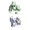
 Basic information
 Components
 Keywords
 Function and homology information
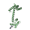
 Homo sapiens (human)
 X-RAY DIFFRACTION /
  Authors
 Canada, 1items
  Citation
 Structure visualization
 Downloads & links
 Other downloads
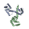


 PDBj
PDBj


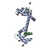
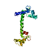
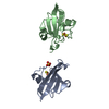
 Assembly
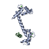


 Mass: 40.078 Da / Num. of mol.: 4 / Source method: obtained synthetically / Formula: Ca
 Mass: 18.015 Da / Num. of mol.: 45 / Source method: isolated from a natural source / Formula: H2O
 Sample preparation
 / Beamline: 23-ID-B / Wavelength: 1.0332 Å
 Processing