Movie
Movie Controller

[English] 日本語
 Yorodumi
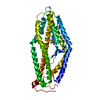
- PDB-6mpv: Cryo-electron microscopy structure of Plasmodium falciparum Rh5/C... -
+ Open data

- Basic information
| Entry | Database: PDB / ID: 6mpv | ||||||
|---|---|---|---|---|---|---|---|
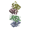
| Title | Cryo-electron microscopy structure of Plasmodium falciparum Rh5/CyRPA/Ripr invasion complex | ||||||
 Components Components |
| ||||||
 Keywords Keywords | CELL INVASION / Invasion Ligand Protein complex | ||||||
| Function / homology |  Function and homology information Function and homology informationmicroneme lumen / microneme / symbiont entry into host / apical part of cell / cytoplasmic vesicle / host extracellular region / host cell plasma membrane / protein-containing complex / extracellular region / plasma membrane Similarity search - Function | ||||||
| Biological species |  | ||||||
| Method | ELECTRON MICROSCOPY / single particle reconstruction / cryo EM / Resolution: 7.17 Å | ||||||
 Authors Authors | Wilson, W. / Zhiheng, Y. / Cowman, A.F. | ||||||
| Funding support |  Australia, 1items Australia, 1items
| ||||||
 Citation Citation | Journal: Nature / Year: 2019 Title: Structure of Plasmodium falciparum Rh5-CyRPA-Ripr invasion complex. Authors: Wilson Wong / Rick Huang / Sebastien Menant / Chuan Hong / Jarrod J Sandow / Richard W Birkinshaw / Julie Healer / Anthony N Hodder / Usheer Kanjee / Christopher J Tonkin / Denise Heckmann / ...Authors: Wilson Wong / Rick Huang / Sebastien Menant / Chuan Hong / Jarrod J Sandow / Richard W Birkinshaw / Julie Healer / Anthony N Hodder / Usheer Kanjee / Christopher J Tonkin / Denise Heckmann / Vladislav Soroka / Teit Max Moscote Søgaard / Thomas Jørgensen / Manoj T Duraisingh / Peter E Czabotar / Willem A de Jongh / Wai-Hong Tham / Andrew I Webb / Zhiheng Yu / Alan F Cowman /   Abstract: Plasmodium falciparum causes the severe form of malaria that has high levels of mortality in humans. Blood-stage merozoites of P. falciparum invade erythrocytes, and this requires interactions ...Plasmodium falciparum causes the severe form of malaria that has high levels of mortality in humans. Blood-stage merozoites of P. falciparum invade erythrocytes, and this requires interactions between multiple ligands from the parasite and receptors in hosts. These interactions include the binding of the Rh5-CyRPA-Ripr complex with the erythrocyte receptor basigin, which is an essential step for entry into human erythrocytes. Here we show that the Rh5-CyRPA-Ripr complex binds the erythrocyte cell line JK-1 significantly better than does Rh5 alone, and that this binding occurs through the insertion of Rh5 and Ripr into host membranes as a complex with high molecular weight. We report a cryo-electron microscopy structure of the Rh5-CyRPA-Ripr complex at subnanometre resolution, which reveals the organization of this essential invasion complex and the mode of interactions between members of the complex, and shows that CyRPA is a critical mediator of complex assembly. Our structure identifies blades 4-6 of the β-propeller of CyRPA as contact sites for Rh5 and Ripr. The limited contacts between Rh5-CyRPA and CyRPA-Ripr are consistent with the dissociation of Rh5 and Ripr from CyRPA for membrane insertion. A comparision of the crystal structure of Rh5-basigin with the cryo-electron microscopy structure of Rh5-CyRPA-Ripr suggests that Rh5 and Ripr are positioned parallel to the erythrocyte membrane before membrane insertion. This provides information on the function of this complex, and thereby provides insights into invasion by P. falciparum. | ||||||
| History |
|
- Structure visualization
| Movie |
Movie viewer |
|---|---|
| Structure viewer | Molecule: MolmilJmol/JSmol |
- Downloads & links
-Download
| PDBx/mmCIF format | 6mpv.cif.gz | 182 KB | Display | PDBx/mmCIF format |
|---|---|---|---|---|
| PDB format | pdb6mpv.ent.gz | 134.7 KB | Display | PDB format |
| PDBx/mmJSON format | 6mpv.json.gz | Tree view | PDBx/mmJSON format | |
| Others |  Other downloads Other downloads |
-Validation report
| Arichive directory | https://data.pdbj.org/pub/pdb/validation_reports/mp/6mpvftp://data.pdbj.org/pub/pdb/validation_reports/mp/6mpv | HTTPS FTP |
|---|
-Related structure data
| Related structure data |  9192MC 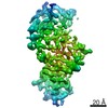 9193C M: map data used to model this data C: citing same article ( |
|---|---|
| Similar structure data |


-Links
 PDBj
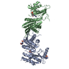
PDBj- Assembly
| Deposited unit | 
|
|---|---|
| 1 |
|
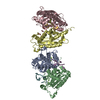
-Components
| #1: Protein | Mass: 39270.195 Da / Num. of mol.: 1 Source method: isolated from a genetically manipulated source Source: (gene. exp.) Strain: isolate 3D7 / Gene: PF3D7_0423800 Production host: Insect cell expression vector pTIE1 (others) References: UniProt: Q8IFM8 |
|---|---|
| #2: Protein | Mass: 39803.371 Da / Num. of mol.: 1 Source method: isolated from a genetically manipulated source Source: (gene. exp.) Production host: Insect cell expression vector pTIE1 (others) References: UniProt: B2L3N7 |
| #3: Protein/peptide | Mass: 1379.692 Da / Num. of mol.: 1 Source method: isolated from a genetically manipulated source Source: (gene. exp.)  |
| #4: Protein/peptide | Mass: 443.539 Da / Num. of mol.: 1 Source method: isolated from a genetically manipulated source Source: (gene. exp.) |
| Has protein modification | Y |
| Sequence details | The sample sequence corresponding to PfRipr is the following: ...The sample sequence corresponding to PfRipr is the following: IDLIEGIFYE |
-Experimental details
-Experiment
| Experiment | Method: ELECTRON MICROSCOPY |
|---|---|
| EM experiment | Aggregation state: PARTICLE / 3D reconstruction method: single particle reconstruction |
- Sample preparation
| Component | Name: PfRh5/CyRPA/PfRipr complex / Type: COMPLEX / Entity ID: all / Source: RECOMBINANT |
|---|---|
| Source (natural) | Organism: |
| Source (recombinant) | Organism: Insect cell expression vector pTIE1 (others) |
| Buffer solution | pH: 8.5 / Details: 20 mM Tris, pH 8.5, 150 mM NaCl |
| Specimen | Embedding applied: NO / Shadowing applied: NO / Staining applied: NO / Vitrification applied: YES |
| Specimen support | Grid material: COPPER / Grid mesh size: 400 divisions/in. / Grid type: Quantifoil R1.2/1.3 |
| Vitrification | Instrument: FEI VITROBOT MARK IV / Cryogen name: ETHANE / Humidity: 100 % / Chamber temperature: 277 K |
- Electron microscopy imaging
| Experimental equipment |  Model: Titan Krios / Image courtesy: FEI Company |
|---|---|
| Microscopy | Model: FEI TITAN KRIOS |
| Electron gun | Electron source:  FIELD EMISSION GUN / Accelerating voltage: 300 kV / Illumination mode: FLOOD BEAM FIELD EMISSION GUN / Accelerating voltage: 300 kV / Illumination mode: FLOOD BEAM |
| Electron lens | Mode: BRIGHT FIELD / Nominal magnification: 105000 X / Calibrated magnification: 48077 X / Cs: 0.01 mm / C2 aperture diameter: 70 µm / Alignment procedure: ZEMLIN TABLEAU |
| Specimen holder | Cryogen: NITROGEN / Specimen holder model: FEI TITAN KRIOS AUTOGRID HOLDER |
| Image recording | Average exposure time: 10 sec. / Electron dose: 92.5 e/Å2 / Detector mode: SUPER-RESOLUTION / Film or detector model: GATAN K2 SUMMIT (4k x 4k) / Num. of grids imaged: 4 / Num. of real images: 12974 |
| EM imaging optics | Energyfilter name: GIF Quantum LS / Energyfilter slit width: 20 eV Spherical aberration corrector: Microscope was equipped with a Cs corrector with two hexapole elements |
| Image scans | Sampling size: 5 µm / Width: 3838 / Height: 3710 / Movie frames/image: 50 |
- Processing
| Software | Name: PHENIX / Version: 1.11_2558: / Classification: refinement | ||||||||||||||||||||||||
|---|---|---|---|---|---|---|---|---|---|---|---|---|---|---|---|---|---|---|---|---|---|---|---|---|---|
| EM software |
| ||||||||||||||||||||||||
| CTF correction | Type: PHASE FLIPPING AND AMPLITUDE CORRECTION | ||||||||||||||||||||||||
| Symmetry | Point symmetry: C1 (asymmetric) | ||||||||||||||||||||||||
| 3D reconstruction | Resolution: 7.17 Å / Resolution method: FSC 0.143 CUT-OFF / Num. of particles: 218837 / Symmetry type: POINT | ||||||||||||||||||||||||
| Atomic model building | Space: REAL | ||||||||||||||||||||||||
| Atomic model building |
| ||||||||||||||||||||||||
| Refine LS restraints |
|