 Movie
Movie Controller

[English] 日本語
 Yorodumi
- PDB-3nh1: Crystal structure of RNase T in complex with a preferred ssDNA (T... -
+ Open data

- Basic information
| Entry | Database: PDB / ID: 3nh1 | ||||||
|---|---|---|---|---|---|---|---|
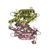
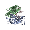
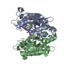
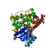
| Title | Crystal structure of RNase T in complex with a preferred ssDNA (TAGG) with two Mg in the active site | ||||||
 Components Components |
| ||||||
 Keywords Keywords | HYDROLASE/DNA / exoribonuclease / RNA processing / RNA maturation / protein-DNA interactions / protein-DNA complex / exo-nuclease / HYDROLASE-DNA complex | ||||||
| Function / homology |  Function and homology information Function and homology informationrRNA 3'-end processing / regulatory ncRNA 3'-end processing / tRNA 3'-end processing / DNA replication proofreading / single-stranded DNA 3'-5' DNA exonuclease activity / 3'-5' exonuclease activity / cellular response to UV / Hydrolases; Acting on ester bonds; Exoribonucleases producing 5'-phosphomonoesters / 3'-5'-RNA exonuclease activity / nucleic acid binding ...rRNA 3'-end processing / regulatory ncRNA 3'-end processing / tRNA 3'-end processing / DNA replication proofreading / single-stranded DNA 3'-5' DNA exonuclease activity / 3'-5' exonuclease activity / cellular response to UV / Hydrolases; Acting on ester bonds; Exoribonucleases producing 5'-phosphomonoesters / 3'-5'-RNA exonuclease activity / nucleic acid binding / DNA damage response / magnesium ion binding / protein homodimerization activity / identical protein binding / cytosol Similarity search - Function | ||||||
| Biological species |  | ||||||
| Method |  X-RAY DIFFRACTION / SYNCHROTRON / MOLECULAR REPLACEMENT / Resolution: 2.107 Å X-RAY DIFFRACTION / SYNCHROTRON / MOLECULAR REPLACEMENT / Resolution: 2.107 Å | ||||||
 Authors Authors | Hsiao, Y.-Y. / Yuan, H.S. | ||||||
 Citation Citation | Journal: Nat.Chem.Biol. / Year: 2011 Title: Structural basis for RNA trimming by RNase T in stable RNA 3'-end maturation Authors: Hsiao, Y.-Y. / Yang, C.-C. / Lin, C.L. / Lin, J.L.J. / Duh, Y. / Yuan, H.S. | ||||||
| History |
|
- Structure visualization
| Structure viewer | Molecule: MolmilJmol/JSmol |
|---|
- Downloads & links
-Download
| PDBx/mmCIF format | 3nh1.cif.gz | 188.1 KB | Display | PDBx/mmCIF format |
|---|---|---|---|---|
| PDB format | pdb3nh1.ent.gz | 146.2 KB | Display | PDB format |
| PDBx/mmJSON format | 3nh1.json.gz | Tree view | PDBx/mmJSON format | |
| Others |  Other downloads Other downloads |
-Validation report
| Arichive directory | https://data.pdbj.org/pub/pdb/validation_reports/nh/3nh1ftp://data.pdbj.org/pub/pdb/validation_reports/nh/3nh1 | HTTPS FTP |
|---|
-Related structure data
| Related structure data | 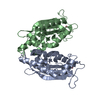 3ngySC 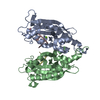 3ngzC 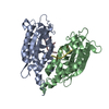 3nh0C 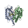 3nh2C S: Starting model for refinement C: citing same article ( |
|---|---|
| Similar structure data |

-Links
 PDBj
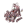
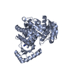
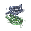
PDBj- Assembly
| Deposited unit | 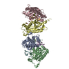
| ||||||||
|---|---|---|---|---|---|---|---|---|---|
| 1 | 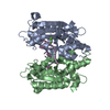
| ||||||||
| 2 | 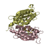
| ||||||||
| Unit cell |
|
-Components
| #1: Protein | Mass: 25719.035 Da / Num. of mol.: 4 Source method: isolated from a genetically manipulated source Source: (gene. exp.) References: UniProt: P30014, Hydrolases; Acting on ester bonds; Exoribonucleases producing 5'-phosphomonoesters #2: DNA chain | Mass: 2152.446 Da / Num. of mol.: 4 / Source method: obtained synthetically / Details: ssDNA #3: Chemical | ChemComp-MG /   Mass: 24.305 Da / Num. of mol.: 8 / Source method: obtained synthetically / Formula: Mg Mass: 24.305 Da / Num. of mol.: 8 / Source method: obtained synthetically / Formula: Mg#4: Water | ChemComp-HOH / |  Mass: 18.015 Da / Num. of mol.: 343 / Source method: isolated from a natural source / Formula: H2O Mass: 18.015 Da / Num. of mol.: 343 / Source method: isolated from a natural source / Formula: H2O |
|---|
-Experimental details
-Experiment
| Experiment | Method: X-RAY DIFFRACTION / Number of used crystals: 1 |
|---|
- Sample preparation
| Crystal | Density Matthews: 1.767152 Å3/Da / Density % sol: 30.396488 % |
|---|---|
| Crystal grow | Temperature: 298 K / Method: vapor diffusion, hanging drop / pH: 6 Details: 0.1M MES monohydrate pH 6.0, 20% w/v PEG MME 2000, VAPOR DIFFUSION, HANGING DROP, temperature 298K |
-Data collection
| Diffraction | Mean temperature: 100 K |
|---|---|
| Diffraction source | Source: SYNCHROTRON / Site: NSRRC  / Beamline: BL13B1 / Wavelength: 0.999 Å / Beamline: BL13B1 / Wavelength: 0.999 Å |
| Detector | Type: ADSC QUANTUM 315 / Detector: CCD / Date: Apr 30, 2010 |
| Radiation | Monochromator: SAGITALLY FOCUSED Si(111) / Protocol: SINGLE WAVELENGTH / Monochromatic (M) / Laue (L): M / Scattering type: x-ray |
| Radiation wavelength | Wavelength: 0.999 Å / Relative weight: 1 |
| Reflection | Resolution: 2.1→30 Å / Num. all: 43799 / Num. obs: 43799 / % possible obs: 99.5 % / Observed criterion σ(F): 0 / Observed criterion σ(I): 0 / Redundancy: 3.2 % / Rsym value: 0.056 / Net I/σ(I): 24.2 |
| Reflection shell | Resolution: 2.1→2.18 Å / Redundancy: 3.1 % / Mean I/σ(I) obs: 4.7 / Num. unique all: 4281 / Rsym value: 0.269 / % possible all: 99.4 |
- Processing
| Software |
| |||||||||||||||||||||||||||||||||||||||||||||||||||||||||||||||||||||||||||||||||||||||||||||||||||||||||||||||||||||||||||||||||||||||||||||||||||||||||||||||||||||||||||||||
|---|---|---|---|---|---|---|---|---|---|---|---|---|---|---|---|---|---|---|---|---|---|---|---|---|---|---|---|---|---|---|---|---|---|---|---|---|---|---|---|---|---|---|---|---|---|---|---|---|---|---|---|---|---|---|---|---|---|---|---|---|---|---|---|---|---|---|---|---|---|---|---|---|---|---|---|---|---|---|---|---|---|---|---|---|---|---|---|---|---|---|---|---|---|---|---|---|---|---|---|---|---|---|---|---|---|---|---|---|---|---|---|---|---|---|---|---|---|---|---|---|---|---|---|---|---|---|---|---|---|---|---|---|---|---|---|---|---|---|---|---|---|---|---|---|---|---|---|---|---|---|---|---|---|---|---|---|---|---|---|---|---|---|---|---|---|---|---|---|---|---|---|---|---|---|---|---|
| Refinement | Method to determine structure: MOLECULAR REPLACEMENT Starting model: PDB ENTRY 3NGY Resolution: 2.107→23.377 Å / Occupancy max: 1 / Occupancy min: 1 / FOM work R set: 0.851 / SU ML: 0.26 / Cross valid method: THROUGHOUT / σ(F): 1.99 / Phase error: 22.87 / Stereochemistry target values: ML
| |||||||||||||||||||||||||||||||||||||||||||||||||||||||||||||||||||||||||||||||||||||||||||||||||||||||||||||||||||||||||||||||||||||||||||||||||||||||||||||||||||||||||||||||
| Solvent computation | Shrinkage radii: 0.9 Å / VDW probe radii: 1.11 Å / Solvent model: FLAT BULK SOLVENT MODEL / Bsol: 36.878 Å2 / ksol: 0.358 e/Å3 | |||||||||||||||||||||||||||||||||||||||||||||||||||||||||||||||||||||||||||||||||||||||||||||||||||||||||||||||||||||||||||||||||||||||||||||||||||||||||||||||||||||||||||||||
| Displacement parameters | Biso max: 86.58 Å2 / Biso mean: 31.0035 Å2 / Biso min: 11.88 Å2
| |||||||||||||||||||||||||||||||||||||||||||||||||||||||||||||||||||||||||||||||||||||||||||||||||||||||||||||||||||||||||||||||||||||||||||||||||||||||||||||||||||||||||||||||
| Refinement step | Cycle: LAST / Resolution: 2.107→23.377 Å
| |||||||||||||||||||||||||||||||||||||||||||||||||||||||||||||||||||||||||||||||||||||||||||||||||||||||||||||||||||||||||||||||||||||||||||||||||||||||||||||||||||||||||||||||
| Refine LS restraints |
| |||||||||||||||||||||||||||||||||||||||||||||||||||||||||||||||||||||||||||||||||||||||||||||||||||||||||||||||||||||||||||||||||||||||||||||||||||||||||||||||||||||||||||||||
| LS refinement shell |
|
