 Movie
Movie Controller

[English] 日本語
 Yorodumi
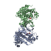
- PDB-1q0h: Crystal structure of selenomethionine-labelled DXR in complex wit... -
+ Open data

- Basic information
| Entry | Database: PDB / ID: 1q0h | ||||||
|---|---|---|---|---|---|---|---|
| Title | Crystal structure of selenomethionine-labelled DXR in complex with fosmidomycin | ||||||
 Components Components | 1-deoxy-D-xylulose 5-phosphate reductoisomerase | ||||||
 Keywords Keywords | OXIDOREDUCTASE | ||||||
| Function / homology |  Function and homology information Function and homology informationDxr protein complex / : / 1-deoxy-D-xylulose-5-phosphate reductoisomerase / 1-deoxy-D-xylulose-5-phosphate reductoisomerase activity / isopentenyl diphosphate biosynthetic process, methylerythritol 4-phosphate pathway / NADPH binding / manganese ion binding / identical protein binding Similarity search - Function | ||||||
| Biological species |  | ||||||
| Method |  X-RAY DIFFRACTION / MOLECULAR REPLACEMENT / Resolution: 2.2 Å X-RAY DIFFRACTION / MOLECULAR REPLACEMENT / Resolution: 2.2 Å | ||||||
 Authors Authors | Mac Sweeney, A. / Lange, R. / D'Arcy, A. / Douangamath, A. / Surivet, J.-P. / Oefner, C. | ||||||
 Citation Citation | Journal: J.Mol.Biol. / Year: 2005 Title: The crystal structure of E.coli 1-deoxy-D-xylulose-5-phosphate reductoisomerase in a ternary complex with the antimalarial compound fosmidomycin and NADPH reveals a tight-binding closed enzyme conformation. Authors: Mac Sweeney, A. / Lange, R. / Fernandes, R.P. / Schulz, H. / Dale, G.E. / Douangamath, A. / Proteau, P.J. / Oefner, C. | ||||||
| History |
|
- Structure visualization
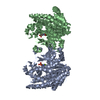
| Structure viewer | Molecule: MolmilJmol/JSmol |
|---|
- Downloads & links
-Download
| PDBx/mmCIF format | 1q0h.cif.gz | 98.6 KB | Display | PDBx/mmCIF format |
|---|---|---|---|---|
| PDB format | pdb1q0h.ent.gz | 74.4 KB | Display | PDB format |
| PDBx/mmJSON format | 1q0h.json.gz | Tree view | PDBx/mmJSON format | |
| Others |  Other downloads Other downloads |
-Validation report
| Arichive directory | https://data.pdbj.org/pub/pdb/validation_reports/q0/1q0hftp://data.pdbj.org/pub/pdb/validation_reports/q0/1q0h | HTTPS FTP |
|---|
-Related structure data
| Related structure data | 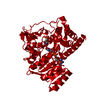 1q0lC  1q0qC  1k5hS C: citing same article ( S: Starting model for refinement |
|---|---|
| Similar structure data |


-Links
 PDBj
PDBj

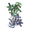
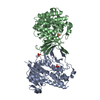
- Assembly
| Deposited unit | 
| ||||||||
|---|---|---|---|---|---|---|---|---|---|
| 1 | 
| ||||||||
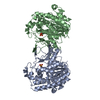
| Unit cell |
| ||||||||
| Details | The second molecule of the biological homodimer is generated by the two-fold axis. Operation x, y, z to y, x, 1-z (non-orthogonal coordinate system) |
-Components
| #1: Protein | Mass: 45295.875 Da / Num. of mol.: 1 Source method: isolated from a genetically manipulated source Source: (gene. exp.) References: UniProt: P45568, 1-deoxy-D-xylulose-5-phosphate reductoisomerase |
|---|---|
| #2: Chemical | ChemComp-FOM / 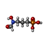  Mass: 183.100 Da / Num. of mol.: 1 / Source method: obtained synthetically / Formula: C4H10NO5P / Comment: antibiotic*YM Mass: 183.100 Da / Num. of mol.: 1 / Source method: obtained synthetically / Formula: C4H10NO5P / Comment: antibiotic*YM |
| #3: Chemical | ChemComp-NDP /   Mass: 745.421 Da / Num. of mol.: 1 / Source method: obtained synthetically / Formula: C21H30N7O17P3 Mass: 745.421 Da / Num. of mol.: 1 / Source method: obtained synthetically / Formula: C21H30N7O17P3 |
| #4: Chemical | ChemComp-CIT /   Mass: 192.124 Da / Num. of mol.: 1 / Source method: obtained synthetically / Formula: C6H8O7 Mass: 192.124 Da / Num. of mol.: 1 / Source method: obtained synthetically / Formula: C6H8O7 |
| #5: Water | ChemComp-HOH /  Mass: 18.015 Da / Num. of mol.: 265 / Source method: isolated from a natural source / Formula: H2O Mass: 18.015 Da / Num. of mol.: 265 / Source method: isolated from a natural source / Formula: H2O |
| Has protein modification | Y |
-Experimental details
-Experiment
| Experiment | Method: X-RAY DIFFRACTION / Number of used crystals: 1 |
|---|
- Sample preparation
| Crystal | Density Matthews: 3.44 Å3/Da / Density % sol: 64.26 % |
|---|---|
| Crystal grow | Temperature: 298 K / Method: vapor diffusion, hanging drop / pH: 5 Details: 2.6M NaCl, 0.1M Acetate-HCl pH 5.0, VAPOR DIFFUSION, HANGING DROP, temperature 298K |
-Data collection
| Diffraction | Mean temperature: 100 K |
|---|---|
| Diffraction source | Source: ROTATING ANODE / Type: ENRAF-NONIUS FR571 / Wavelength: 1.5418 Å |
| Detector | Type: MARRESEARCH / Detector: IMAGE PLATE / Date: Mar 1, 2003 / Details: Osmic mirrors |
| Radiation | Protocol: SINGLE WAVELENGTH / Monochromatic (M) / Laue (L): M / Scattering type: x-ray |
| Radiation wavelength | Wavelength: 1.5418 Å / Relative weight: 1 |
| Reflection | Resolution: 2.2→20 Å / Num. all: 32094 / Num. obs: 31221 / % possible obs: 97.1 % / Observed criterion σ(F): 6 / Observed criterion σ(I): 6 / Redundancy: 3.8 % / Biso Wilson estimate: 0.23 Å2 / Rmerge(I) obs: 0.129 / Net I/σ(I): 7.64 |
| Reflection shell | Resolution: 2.2→2.34 Å / Rmerge(I) obs: 0.465 / Mean I/σ(I) obs: 1.97 / % possible all: 94.5 |
- Processing
| Software |
| ||||||||||||||||||||
|---|---|---|---|---|---|---|---|---|---|---|---|---|---|---|---|---|---|---|---|---|---|
| Refinement | Method to determine structure: MOLECULAR REPLACEMENT Starting model: PDB ENTRY 1K5H (NADPH binding domain) Resolution: 2.2→17.62 Å / Isotropic thermal model: isotropic / σ(F): 6 / Stereochemistry target values: Engh & Huber
| ||||||||||||||||||||
| Displacement parameters | Biso mean: 23.8 Å2 | ||||||||||||||||||||
| Refinement step | Cycle: LAST / Resolution: 2.2→17.62 Å
| ||||||||||||||||||||
| Refine LS restraints |
|
