 Movie
Movie Controller


+ Open data

- Basic information
| Entry | Database: PDB / ID: 5di0 | ||||||
|---|---|---|---|---|---|---|---|
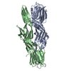
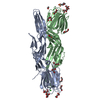
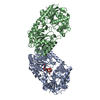
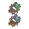
| Title | Crystal structure of Dln1 | ||||||
 Components Components | Natterin-like protein | ||||||
 Keywords Keywords | SUGAR BINDING PROTEIN / Pore-forming protein / Aeolysin-like protein / Vetebrate / High-mannose glycans / Complex | ||||||
| Function / homology |  Function and homology information Function and homology informationdisaccharide binding / pore complex / D-mannose binding / defense response to bacterium / identical protein binding / plasma membrane Similarity search - Function | ||||||
| Biological species |  | ||||||
| Method |  X-RAY DIFFRACTION / SYNCHROTRON / MOLECULAR REPLACEMENT / Resolution: 1.7 Å X-RAY DIFFRACTION / SYNCHROTRON / MOLECULAR REPLACEMENT / Resolution: 1.7 Å | ||||||
 Authors Authors | Jia, N. / Jiang, Y.L. / Cheng, W. / Wang, H.W. / Zhou, C.Z. / Chen, Y. | ||||||
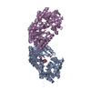
 Citation Citation | Journal: EMBO Rep / Year: 2016 Title: Structural basis for receptor recognition and pore formation of a zebrafish aerolysin-like protein. Authors: Ning Jia / Nan Liu / Wang Cheng / Yong-Liang Jiang / Hui Sun / Lan-Lan Chen / Junhui Peng / Yonghui Zhang / Yue-He Ding / Zhi-Hui Zhang / Xuejuan Wang / Gang Cai / Junfeng Wang / Meng-Qiu ...Authors: Ning Jia / Nan Liu / Wang Cheng / Yong-Liang Jiang / Hui Sun / Lan-Lan Chen / Junhui Peng / Yonghui Zhang / Yue-He Ding / Zhi-Hui Zhang / Xuejuan Wang / Gang Cai / Junfeng Wang / Meng-Qiu Dong / Zhiyong Zhang / Hui Wu / Hong-Wei Wang / Yuxing Chen / Cong-Zhao Zhou /   Abstract: Various aerolysin-like pore-forming proteins have been identified from bacteria to vertebrates. However, the mechanism of receptor recognition and/or pore formation of the eukaryotic members remains ...Various aerolysin-like pore-forming proteins have been identified from bacteria to vertebrates. However, the mechanism of receptor recognition and/or pore formation of the eukaryotic members remains unknown. Here, we present the first crystal and electron microscopy structures of a vertebrate aerolysin-like protein from Danio rerio, termed Dln1, before and after pore formation. Each subunit of Dln1 dimer comprises a β-prism lectin module followed by an aerolysin module. Specific binding of the lectin module toward high-mannose glycans triggers drastic conformational changes of the aerolysin module in a pH-dependent manner, ultimately resulting in the formation of a membrane-bound octameric pore. Structural analyses combined with computational simulations and biochemical assays suggest a pore-forming process with an activation mechanism distinct from the previously characterized bacterial members. Moreover, Dln1 and its homologs are ubiquitously distributed in bony fishes and lamprey, suggesting a novel fish-specific defense molecule. | ||||||
| History |
|
- Structure visualization
| Structure viewer | Molecule: MolmilJmol/JSmol |
|---|
- Downloads & links
-Download
| PDBx/mmCIF format | 5di0.cif.gz | 166.3 KB | Display | PDBx/mmCIF format |
|---|---|---|---|---|
| PDB format | pdb5di0.ent.gz | 128.3 KB | Display | PDB format |
| PDBx/mmJSON format | 5di0.json.gz | Tree view | PDBx/mmJSON format | |
| Others |  Other downloads Other downloads |
-Validation report
| Arichive directory | https://data.pdbj.org/pub/pdb/validation_reports/di/5di0ftp://data.pdbj.org/pub/pdb/validation_reports/di/5di0 | HTTPS FTP |
|---|
-Related structure data
| Related structure data | 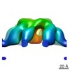 3244C  4znoSC  4znqC  4znrC S: Starting model for refinement C: citing same article ( |
|---|---|
| Similar structure data |


-Links
 PDBj
PDBj- Assembly
| Deposited unit | 
| ||||||||
|---|---|---|---|---|---|---|---|---|---|
| 1 |
| ||||||||
| Unit cell |
|
-Components
-Protein , 1 types, 2 molecules AB
| #1: Protein | Mass: 36478.879 Da / Num. of mol.: 2 Source method: isolated from a genetically manipulated source Source: (gene. exp.)  |
|---|
-Non-polymers , 6 types, 887 molecules 


| #2: Chemical | ChemComp-EDO /  Mass: 62.068 Da / Num. of mol.: 12 / Source method: obtained synthetically / Formula: C2H6O2 Mass: 62.068 Da / Num. of mol.: 12 / Source method: obtained synthetically / Formula: C2H6O2#3: Chemical | ChemComp-PEG /  Mass: 106.120 Da / Num. of mol.: 8 / Source method: obtained synthetically / Formula: C4H10O3 Mass: 106.120 Da / Num. of mol.: 8 / Source method: obtained synthetically / Formula: C4H10O3#4: Chemical | ChemComp-PGE /  Mass: 150.173 Da / Num. of mol.: 7 / Source method: obtained synthetically / Formula: C6H14O4 Mass: 150.173 Da / Num. of mol.: 7 / Source method: obtained synthetically / Formula: C6H14O4#5: Chemical | ChemComp-PG4 /  Mass: 194.226 Da / Num. of mol.: 6 / Source method: obtained synthetically / Formula: C8H18O5 / Comment: precipitant*YM Mass: 194.226 Da / Num. of mol.: 6 / Source method: obtained synthetically / Formula: C8H18O5 / Comment: precipitant*YM#6: Chemical |  Mass: 35.453 Da / Num. of mol.: 2 / Source method: obtained synthetically / Formula: Cl Mass: 35.453 Da / Num. of mol.: 2 / Source method: obtained synthetically / Formula: Cl#7: Water | ChemComp-HOH / | Mass: 18.015 Da / Num. of mol.: 852 / Source method: isolated from a natural source / Formula: H2O |
|---|
-Experimental details
-Experiment
| Experiment | Method: X-RAY DIFFRACTION |
|---|
- Sample preparation
| Crystal | Density Matthews: 2.86 Å3/Da / Density % sol: 57.1 % |
|---|---|
| Crystal grow | Temperature: 289 K / Method: vapor diffusion / pH: 4 / Details: 12% PEG 4K,0.2M NH4Ac, 0.1M NaAc pH 4.0 |
-Data collection
| Diffraction | Mean temperature: 100 K |
|---|---|
| Diffraction source | Source: SYNCHROTRON / Site: SSRF / Beamline: BL17U / Wavelength: 0.97911 Å |
| Detector | Type: ADSC QUANTUM 315r / Detector: CCD / Date: Mar 3, 2015 |
| Radiation | Protocol: SINGLE WAVELENGTH / Monochromatic (M) / Laue (L): M / Scattering type: x-ray |
| Radiation wavelength | Wavelength: 0.97911 Å / Relative weight: 1 |
| Reflection | Resolution: 1.7→50 Å / Num. obs: 87589 / % possible obs: 98.6 % / Redundancy: 3.4 % / Net I/σ(I): 14.8 |
| Reflection shell | Resolution: 1.7→1.76 Å / Redundancy: 3.4 % / Rmerge(I) obs: 0.459 / Mean I/σ(I) obs: 2.9 / % possible all: 98.7 |
- Processing
| Software |
| ||||||||||||||||||||||||||||||||||||||||||||||||||||||||||||||||||||||||||||||||||||||||||||||||||||||||||||||||||||||||||||||||||||||||||||||||||||||||||||||||||||||||||||||||||||||
|---|---|---|---|---|---|---|---|---|---|---|---|---|---|---|---|---|---|---|---|---|---|---|---|---|---|---|---|---|---|---|---|---|---|---|---|---|---|---|---|---|---|---|---|---|---|---|---|---|---|---|---|---|---|---|---|---|---|---|---|---|---|---|---|---|---|---|---|---|---|---|---|---|---|---|---|---|---|---|---|---|---|---|---|---|---|---|---|---|---|---|---|---|---|---|---|---|---|---|---|---|---|---|---|---|---|---|---|---|---|---|---|---|---|---|---|---|---|---|---|---|---|---|---|---|---|---|---|---|---|---|---|---|---|---|---|---|---|---|---|---|---|---|---|---|---|---|---|---|---|---|---|---|---|---|---|---|---|---|---|---|---|---|---|---|---|---|---|---|---|---|---|---|---|---|---|---|---|---|---|---|---|---|---|
| Refinement | Method to determine structure: MOLECULAR REPLACEMENT Starting model: 4ZNO Resolution: 1.7→47.6 Å / Cor.coef. Fo:Fc: 0.967 / Cor.coef. Fo:Fc free: 0.951 / SU B: 1.918 / SU ML: 0.064 / Cross valid method: THROUGHOUT / ESU R: 0.096 / ESU R Free: 0.098 / Stereochemistry target values: MAXIMUM LIKELIHOOD / Details: HYDROGENS HAVE BEEN ADDED IN THE RIDING POSITIONS
| ||||||||||||||||||||||||||||||||||||||||||||||||||||||||||||||||||||||||||||||||||||||||||||||||||||||||||||||||||||||||||||||||||||||||||||||||||||||||||||||||||||||||||||||||||||||
| Solvent computation | Ion probe radii: 0.8 Å / Shrinkage radii: 0.8 Å / VDW probe radii: 1.2 Å / Solvent model: MASK | ||||||||||||||||||||||||||||||||||||||||||||||||||||||||||||||||||||||||||||||||||||||||||||||||||||||||||||||||||||||||||||||||||||||||||||||||||||||||||||||||||||||||||||||||||||||
| Displacement parameters | Biso mean: 27.158 Å2
| ||||||||||||||||||||||||||||||||||||||||||||||||||||||||||||||||||||||||||||||||||||||||||||||||||||||||||||||||||||||||||||||||||||||||||||||||||||||||||||||||||||||||||||||||||||||
| Refinement step | Cycle: 1 / Resolution: 1.7→47.6 Å
| ||||||||||||||||||||||||||||||||||||||||||||||||||||||||||||||||||||||||||||||||||||||||||||||||||||||||||||||||||||||||||||||||||||||||||||||||||||||||||||||||||||||||||||||||||||||
| Refine LS restraints |
|