 Movie
Movie Controller


+ Open data

- Basic information
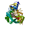
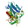
| Entry | Database: PDB / ID: 3f5o | ||||||
|---|---|---|---|---|---|---|---|
| Title | Crystal Structure of hTHEM2(undecan-2-one-CoA) complex | ||||||
 Components Components | Thioesterase superfamily member 2 | ||||||
 Keywords Keywords | HYDROLASE / hotdog fold | ||||||
| Function / homology |  Function and homology information Function and homology informationpalmitoyl-CoA hydrolase / fatty acyl-CoA hydrolase activity / Mitochondrial Fatty Acid Beta-Oxidation / Hydrolases; Acting on ester bonds; Thioester hydrolases / negative regulation of cold-induced thermogenesis / lipid metabolic process / spindle / protein homotetramerization / mitochondrial matrix / mitochondrion ...palmitoyl-CoA hydrolase / fatty acyl-CoA hydrolase activity / Mitochondrial Fatty Acid Beta-Oxidation / Hydrolases; Acting on ester bonds; Thioester hydrolases / negative regulation of cold-induced thermogenesis / lipid metabolic process / spindle / protein homotetramerization / mitochondrial matrix / mitochondrion / metal ion binding / nucleus / cytosol Similarity search - Function | ||||||
| Biological species |  Homo sapiens (human) Homo sapiens (human) | ||||||
| Method |  X-RAY DIFFRACTION / MOLECULAR REPLACEMENT / Resolution: 1.7 Å X-RAY DIFFRACTION / MOLECULAR REPLACEMENT / Resolution: 1.7 Å | ||||||
 Authors Authors | Xu, H. / Gong, W. | ||||||
 Citation Citation | Journal: Biochemistry / Year: 2009 Title: The mechanisms of human hotdog-fold thioesterase 2 (hTHEM2) substrate recognition and catalysis illuminated by a structure and function based analysis Authors: Cao, J. / Xu, H. / Zhao, H. / Gong, W. / Dunaway-Mariano, D. | ||||||
| History |
|
- Structure visualization
| Structure viewer | Molecule: MolmilJmol/JSmol |
|---|
- Downloads & links
-Download
| PDBx/mmCIF format | 3f5o.cif.gz | 247 KB | Display | PDBx/mmCIF format |
|---|---|---|---|---|
| PDB format | pdb3f5o.ent.gz | 199.2 KB | Display | PDB format |
| PDBx/mmJSON format | 3f5o.json.gz | Tree view | PDBx/mmJSON format | |
| Others |  Other downloads Other downloads |
-Validation report
| Arichive directory | https://data.pdbj.org/pub/pdb/validation_reports/f5/3f5oftp://data.pdbj.org/pub/pdb/validation_reports/f5/3f5o | HTTPS FTP |
|---|
-Related structure data
| Related structure data |  2f0xS S: Starting model for refinement |
|---|---|
| Similar structure data |


-Links
 PDBj
PDBj


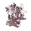
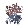
- Assembly
| Deposited unit | 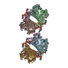
| ||||||||
|---|---|---|---|---|---|---|---|---|---|
| 1 | 
| ||||||||
| 2 | 
| ||||||||
| Unit cell |
|
-Components
-Protein , 1 types, 8 molecules ABCDEFGH
| #1: Protein | Mass: 16052.646 Da / Num. of mol.: 8 Source method: isolated from a genetically manipulated source Source: (gene. exp.) Homo sapiens (human) / Plasmid: pET22b / Production host:  |
|---|
-Non-polymers , 5 types, 877 molecules 


| #2: Chemical | ChemComp-CL /  Mass: 35.453 Da / Num. of mol.: 7 / Source method: obtained synthetically / Formula: Cl Mass: 35.453 Da / Num. of mol.: 7 / Source method: obtained synthetically / Formula: Cl#3: Chemical | ChemComp-UOC /  Mass: 170.292 Da / Num. of mol.: 8 / Source method: obtained synthetically / Formula: C11H22O Mass: 170.292 Da / Num. of mol.: 8 / Source method: obtained synthetically / Formula: C11H22O#4: Chemical | ChemComp-COA /  Mass: 767.534 Da / Num. of mol.: 8 / Source method: obtained synthetically / Formula: C21H36N7O16P3S Mass: 767.534 Da / Num. of mol.: 8 / Source method: obtained synthetically / Formula: C21H36N7O16P3S#5: Chemical | ChemComp-P6G /  Mass: 282.331 Da / Num. of mol.: 4 / Source method: obtained synthetically / Formula: C12H26O7 / Comment: precipitant*YM Mass: 282.331 Da / Num. of mol.: 4 / Source method: obtained synthetically / Formula: C12H26O7 / Comment: precipitant*YM#6: Water | ChemComp-HOH / | Mass: 18.015 Da / Num. of mol.: 850 / Source method: isolated from a natural source / Formula: H2O |
|---|
-Experimental details
-Experiment
| Experiment | Method: X-RAY DIFFRACTION / Number of used crystals: 1 |
|---|
- Sample preparation
| Crystal | Density Matthews: 2.35 Å3/Da / Density % sol: 47.64 % |
|---|---|
| Crystal grow | Temperature: 293 K / Method: vapor diffusion, hanging drop / pH: 7.5 Details: 15% PEG3350, 0.1M ammonium citrate, dibasic, 5-10% glycerol, pH 7.5, VAPOR DIFFUSION, HANGING DROP, temperature 293K |
-Data collection
| Diffraction | Mean temperature: 100 K |
|---|---|
| Diffraction source | Source: ROTATING ANODE / Type: RIGAKU FR-E+ SUPERBRIGHT / Wavelength: 1.5418 Å |
| Detector | Type: RIGAKU RAXIS IV++ / Detector: IMAGE PLATE / Date: Nov 19, 2007 |
| Radiation | Protocol: SINGLE WAVELENGTH / Monochromatic (M) / Laue (L): M / Scattering type: x-ray |
| Radiation wavelength | Wavelength: 1.5418 Å / Relative weight: 1 |
| Reflection | Resolution: 1.7→50 Å / Num. obs: 134034 / % possible obs: 99.1 % |
| Reflection shell | Resolution: 1.7→1.76 Å / % possible all: 99.2 |
- Processing
| Software |
| ||||||||||||||||||||||||||||||||||||||||||||||||||||||||||||||||||||||||||||||||||||||||||
|---|---|---|---|---|---|---|---|---|---|---|---|---|---|---|---|---|---|---|---|---|---|---|---|---|---|---|---|---|---|---|---|---|---|---|---|---|---|---|---|---|---|---|---|---|---|---|---|---|---|---|---|---|---|---|---|---|---|---|---|---|---|---|---|---|---|---|---|---|---|---|---|---|---|---|---|---|---|---|---|---|---|---|---|---|---|---|---|---|---|---|---|
| Refinement | Method to determine structure: MOLECULAR REPLACEMENT Starting model: 2F0X Resolution: 1.7→20.73 Å / Cor.coef. Fo:Fc: 0.966 / Cor.coef. Fo:Fc free: 0.95 / Cross valid method: THROUGHOUT / ESU R: 0.114 / ESU R Free: 0.113 / Stereochemistry target values: MAXIMUM LIKELIHOOD / Details: HYDROGENS HAVE BEEN ADDED IN THE RIDING POSITIONS
| ||||||||||||||||||||||||||||||||||||||||||||||||||||||||||||||||||||||||||||||||||||||||||
| Solvent computation | Ion probe radii: 0.8 Å / Shrinkage radii: 0.8 Å / VDW probe radii: 1.2 Å / Solvent model: MASK | ||||||||||||||||||||||||||||||||||||||||||||||||||||||||||||||||||||||||||||||||||||||||||
| Displacement parameters | Biso mean: 27.255 Å2
| ||||||||||||||||||||||||||||||||||||||||||||||||||||||||||||||||||||||||||||||||||||||||||
| Refinement step | Cycle: LAST / Resolution: 1.7→20.73 Å
| ||||||||||||||||||||||||||||||||||||||||||||||||||||||||||||||||||||||||||||||||||||||||||
| Refine LS restraints |
| ||||||||||||||||||||||||||||||||||||||||||||||||||||||||||||||||||||||||||||||||||||||||||
| LS refinement shell | Resolution: 1.696→1.74 Å / Total num. of bins used: 20
|
