 Movie
Movie Controller
 Structure viewers
 About Yorodumi Papers


+Search query
-Structure paper
| Title | Structural basis for receptor recognition and pore formation of a zebrafish aerolysin-like protein. |
|---|---|
| Journal, issue, pages | EMBO Rep, Vol. 17, Issue 2, Page 235-248, Year 2016 |
| Publish date | Dec 28, 2015 |
 Authors Authors | Ning Jia / Nan Liu / Wang Cheng / Yong-Liang Jiang / Hui Sun / Lan-Lan Chen / Junhui Peng / Yonghui Zhang / Yue-He Ding / Zhi-Hui Zhang / Xuejuan Wang / Gang Cai / Junfeng Wang / Meng-Qiu Dong / Zhiyong Zhang / Hui Wu / Hong-Wei Wang / Yuxing Chen / Cong-Zhao Zhou /   |
| PubMed Abstract | Various aerolysin-like pore-forming proteins have been identified from bacteria to vertebrates. However, the mechanism of receptor recognition and/or pore formation of the eukaryotic members remains ...Various aerolysin-like pore-forming proteins have been identified from bacteria to vertebrates. However, the mechanism of receptor recognition and/or pore formation of the eukaryotic members remains unknown. Here, we present the first crystal and electron microscopy structures of a vertebrate aerolysin-like protein from Danio rerio, termed Dln1, before and after pore formation. Each subunit of Dln1 dimer comprises a β-prism lectin module followed by an aerolysin module. Specific binding of the lectin module toward high-mannose glycans triggers drastic conformational changes of the aerolysin module in a pH-dependent manner, ultimately resulting in the formation of a membrane-bound octameric pore. Structural analyses combined with computational simulations and biochemical assays suggest a pore-forming process with an activation mechanism distinct from the previously characterized bacterial members. Moreover, Dln1 and its homologs are ubiquitously distributed in bony fishes and lamprey, suggesting a novel fish-specific defense molecule. |
 External links External links | EMBO Rep / PubMed:26711430 / PubMed Central |
| Methods | EM (electron crystallography) / X-ray diffraction |
| Resolution | 1.7 - 20.0 Å |
| Structure data | 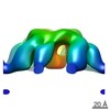 EMDB-3244:  PDB-4zno:  PDB-4znq:  PDB-4znr:  PDB-5di0: |
| Chemicals |  ChemComp-CL:  ChemComp-EPE:  ChemComp-HOH:  ChemComp-PEG:  ChemComp-PGE:  ChemComp-PG4:  ChemComp-EDO:  ChemComp-1PE: |
| Source |
|
 Keywords Keywords | SUGAR BINDING PROTEIN / Pore-forming protein / Aeolysin-like protein / Vetebrate / High-mannose glycans / Complex |

