 Movie
Movie Controller

[English] 日本語
 Yorodumi
- PDB-6mk9: X-ray crystal structure of darunavir-resistant-P51 HIV-1 protease... -
+ Open data

- Basic information
| Entry | Database: PDB / ID: 6mk9 | |||||||||
|---|---|---|---|---|---|---|---|---|---|---|
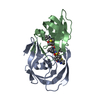
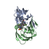
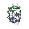
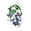
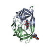
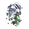
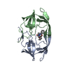
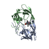
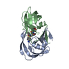
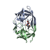
| Title | X-ray crystal structure of darunavir-resistant-P51 HIV-1 protease in complex with GRL-121 | |||||||||
 Components Components | Protease | |||||||||
 Keywords Keywords | HYDROLASE/HYDROLASE INHIBITOR / protease-inhibitor complex / darunavir-resistance / P51 / GRL-121 / non-peptidic / HYDROLASE / HYDROLASE-HYDROLASE INHIBITOR complex | |||||||||
| Function / homology |  Function and homology information Function and homology informationhost multivesicular body / aspartic-type endopeptidase activity / virion membrane / proteolysis Similarity search - Function | |||||||||
| Biological species |   Human immunodeficiency virus 1 Human immunodeficiency virus 1 | |||||||||
| Method |  X-RAY DIFFRACTION / SYNCHROTRON / MOLECULAR REPLACEMENT / Resolution: 1.7 Å X-RAY DIFFRACTION / SYNCHROTRON / MOLECULAR REPLACEMENT / Resolution: 1.7 Å | |||||||||
 Authors Authors | Yedidi, R.S. / Hayashi, H. / Das, D. / Mitsuya, H. | |||||||||
| Funding support |  United States, 1items United States, 1items
| |||||||||
 Citation Citation | Journal: To Be Published Title: X-ray crystal structure of darunavir-resistant-P51 HIV-1 protease in complex with GRL-121 Authors: Yedidi, R.S. / Hayashi, H. / Das, D. / Mitsuya, H. #1: Journal: J. Virol. / Year: 2010 Title: In vitro selection of highly darunavir-resistant and replication-competent HIV-1 variants by using a mixture of clinical HIV-1 isolates resistant to multiple conventional protease inhibitors. Authors: Koh, Y. / Amano, M. / Towata, T. / Danish, M. / Leshchenko-Yashchuk, S. / Das, D. / Nakayama, M. / Tojo, Y. / Ghosh, A.K. / Mitsuya, H. | |||||||||
| History |
|
- Structure visualization
| Structure viewer | Molecule: MolmilJmol/JSmol |
|---|
- Downloads & links
-Download
| PDBx/mmCIF format | 6mk9.cif.gz | 60.3 KB | Display | PDBx/mmCIF format |
|---|---|---|---|---|
| PDB format | pdb6mk9.ent.gz | 42.9 KB | Display | PDB format |
| PDBx/mmJSON format | 6mk9.json.gz | Tree view | PDBx/mmJSON format | |
| Others |  Other downloads Other downloads |
-Validation report
| Arichive directory | https://data.pdbj.org/pub/pdb/validation_reports/mk/6mk9ftp://data.pdbj.org/pub/pdb/validation_reports/mk/6mk9 | HTTPS FTP |
|---|
-Related structure data
| Related structure data |  4hlaS S: Starting model for refinement |
|---|---|
| Similar structure data |

-Links
 PDBj
PDBj
- Assembly
| Deposited unit | 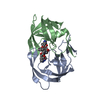
| ||||||||
|---|---|---|---|---|---|---|---|---|---|
| 1 |
| ||||||||
| Unit cell |
|
-Components
| #1: Protein | Mass: 10875.717 Da / Num. of mol.: 2 Source method: isolated from a genetically manipulated source Source: (gene. exp.) Human immunodeficiency virus 1 / Gene: pol / Production host:  #2: Chemical | ChemComp-7O7 / ( |   Mass: 670.839 Da / Num. of mol.: 1 / Source method: obtained synthetically / Formula: C33H42N4O7S2 Mass: 670.839 Da / Num. of mol.: 1 / Source method: obtained synthetically / Formula: C33H42N4O7S2#3: Water | ChemComp-HOH / |  Mass: 18.015 Da / Num. of mol.: 212 / Source method: isolated from a natural source / Formula: H2O Mass: 18.015 Da / Num. of mol.: 212 / Source method: isolated from a natural source / Formula: H2OHas protein modification | N | |
|---|
-Experimental details
-Experiment
| Experiment | Method: X-RAY DIFFRACTION / Number of used crystals: 1 |
|---|
- Sample preparation
| Crystal | Density Matthews: 2.17 Å3/Da / Density % sol: 54.36 % |
|---|---|
| Crystal grow | Temperature: 298 K / Method: vapor diffusion, hanging drop / pH: 6.8 Details: 0.15 M Ammonium sulfate o.1 M HEPES pH6.8 15% (W/V) PEG 4000 |
-Data collection
| Diffraction | Mean temperature: 100 K |
|---|---|
| Diffraction source | Source: SYNCHROTRON / Site: SPring-8  / Beamline: BL41XU / Wavelength: 1 Å / Beamline: BL41XU / Wavelength: 1 Å |
| Detector | Type: DECTRIS PILATUS3 6M / Detector: PIXEL / Date: Jul 16, 2014 |
| Radiation | Protocol: SINGLE WAVELENGTH / Monochromatic (M) / Laue (L): M / Scattering type: x-ray |
| Radiation wavelength | Wavelength: 1 Å / Relative weight: 1 |
| Reflection | Resolution: 1.6→50 Å / Num. obs: 24079 / % possible obs: 99.3 % / Redundancy: 9.4 % / Rmerge(I) obs: 0.08 / Net I/σ(I): 26.05 |
| Reflection shell | Resolution: 1.6→1.63 Å / Redundancy: 9 % / Rmerge(I) obs: 0.57 / Mean I/σ(I) obs: 3.1 / Num. unique obs: 1177 / % possible all: 98.9 |
- Processing
| Software |
| ||||||||||||||||||||||||||||||||||||||||||||||||||||||||
|---|---|---|---|---|---|---|---|---|---|---|---|---|---|---|---|---|---|---|---|---|---|---|---|---|---|---|---|---|---|---|---|---|---|---|---|---|---|---|---|---|---|---|---|---|---|---|---|---|---|---|---|---|---|---|---|---|---|
| Refinement | Method to determine structure: MOLECULAR REPLACEMENT Starting model: 4HLA Resolution: 1.7→32.943 Å / SU ML: 0.18 / Cross valid method: FREE R-VALUE / σ(F): 0.23 / Phase error: 19.82
| ||||||||||||||||||||||||||||||||||||||||||||||||||||||||
| Solvent computation | Shrinkage radii: 0.9 Å / VDW probe radii: 1.11 Å | ||||||||||||||||||||||||||||||||||||||||||||||||||||||||
| Refinement step | Cycle: LAST / Resolution: 1.7→32.943 Å
| ||||||||||||||||||||||||||||||||||||||||||||||||||||||||
| Refine LS restraints |
| ||||||||||||||||||||||||||||||||||||||||||||||||||||||||
| LS refinement shell | Refine-ID: X-RAY DIFFRACTION
|
