 ムービー
ムービー コントローラー


+ データを開く

- 基本情報
基本情報
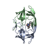
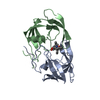
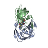
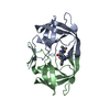
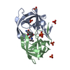
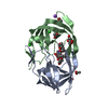
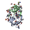
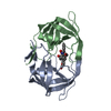
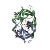
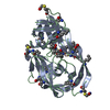
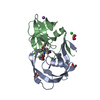
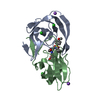
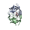
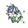
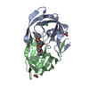
| 登録情報 | データベース: PDB / ID: 1kzk | ||||||
|---|---|---|---|---|---|---|---|
| タイトル | JE-2147-HIV Protease Complex | ||||||
 要素 要素 | Protease | ||||||
 キーワード キーワード | HYDROLASE/HYDROLASE INHIBITOR / HIV Protease Complex / Anisotropic Displacement Parameters / Viral protein / hydrolase / HYDROLASE-HYDROLASE INHIBITOR complex | ||||||
| 機能・相同性 |  機能・相同性情報 機能・相同性情報HIV-1 retropepsin / symbiont-mediated activation of host apoptosis / retroviral ribonuclease H / exoribonuclease H / exoribonuclease H activity / DNA integration / viral genome integration into host DNA / establishment of integrated proviral latency / RNA-directed DNA polymerase / RNA stem-loop binding ...HIV-1 retropepsin / symbiont-mediated activation of host apoptosis / retroviral ribonuclease H / exoribonuclease H / exoribonuclease H activity / DNA integration / viral genome integration into host DNA / establishment of integrated proviral latency / RNA-directed DNA polymerase / RNA stem-loop binding / viral penetration into host nucleus / host multivesicular body / RNA-directed DNA polymerase activity / RNA-DNA hybrid ribonuclease activity / 転移酵素; リンを含む基を移すもの; 核酸を移すもの / host cell / viral nucleocapsid / DNA recombination / DNA-directed DNA polymerase / aspartic-type endopeptidase activity / 加水分解酵素; エステル加水分解酵素 / DNA-directed DNA polymerase activity / symbiont-mediated suppression of host gene expression / viral translational frameshifting / symbiont entry into host cell / lipid binding / host cell nucleus / host cell plasma membrane / virion membrane / structural molecule activity / proteolysis / DNA binding / zinc ion binding 類似検索 - 分子機能 | ||||||
| 生物種 |   Human immunodeficiency virus (ヒト免疫不全ウイルス) Human immunodeficiency virus (ヒト免疫不全ウイルス) | ||||||
| 手法 |  X線回折 / シンクロトロン / 分子置換 / 解像度: 1.09 Å X線回折 / シンクロトロン / 分子置換 / 解像度: 1.09 Å | ||||||
 データ登録者 データ登録者 | Reiling, K.K. / Endres, N.F. / Dauber, D.S. / Craik, C.S. / Stroud, R.M. | ||||||
 引用 引用 | ジャーナル: Biochemistry / 年: 2002 タイトル: Anisotropic Dynamics of the JE-2147-HIV Protease Complex: Drug Resistance and Thermodynamic Binding Mode Examined in a 1.09 A Structure 著者: Reiling, K.K. / Endres, N.F. / Dauber, D.S. / Craik, C.S. / Stroud, R.M. #1: ジャーナル: J.Med.Chem. / 年: 1999タイトル: Structure-Activity Relationship of Small-Sized HIV Protease Inhibitors Containing Allophenylnorstatine 著者: Mimoto, T. / Kato, R. / Takaku, H. / Nojima, S. / Terashima, K. / Misawa, S. / Fukazawa, T. / Ueno, T. / Sato, H. / Shintani, M. / Kiso, Y. / Hayashi, H. | ||||||
| 履歴 |
|
- 構造の表示
構造の表示
| 構造ビューア | 分子: MolmilJmol/JSmol |
|---|
- ダウンロードとリンク
-ダウンロード
| PDBx/mmCIF形式 | 1kzk.cif.gz | 137.4 KB | 表示 | PDBx/mmCIF形式 |
|---|---|---|---|---|
| PDB形式 | pdb1kzk.ent.gz | 107.6 KB | 表示 | PDB形式 |
| PDBx/mmJSON形式 | 1kzk.json.gz | ツリー表示 | PDBx/mmJSON形式 | |
| その他 |  その他のダウンロード その他のダウンロード |
-検証レポート
| アーカイブディレクトリ | https://data.pdbj.org/pub/pdb/validation_reports/kz/1kzkftp://data.pdbj.org/pub/pdb/validation_reports/kz/1kzk | HTTPS FTP |
|---|
-関連構造データ
| 関連構造データ | 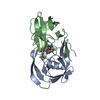 1hpxS S: 精密化の開始モデル |
|---|---|
| 類似構造データ |


-リンク
 PDBj
PDBj

- 集合体
集合体
| 登録構造単位 | 
| ||||||||
|---|---|---|---|---|---|---|---|---|---|
| 1 |
| ||||||||
| 単位格子 |
| ||||||||
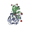
| 詳細 | The Protease is active as a Dimer |
-要素
| #1: タンパク質 | 分子量: 10801.763 Da / 分子数: 2 / 変異: Q7K, K14R, S37N, R41K, L63P, I64V / 由来タイプ: 組換発現 由来: (組換発現) Human immunodeficiency virus (ヒト免疫不全ウイルス)属: Lentivirus / プラスミド: pTacTac / 発現宿主:  #2: 化合物 | ChemComp-JE2 / ( | 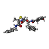  タイプ: peptide-like, Peptide-like / クラス: 酵素阻害剤 / 分子量: 575.718 Da / 分子数: 1 / 由来タイプ: 合成 / 式: C32H37N3O5S / 参照: KNI-764 タイプ: peptide-like, Peptide-like / クラス: 酵素阻害剤 / 分子量: 575.718 Da / 分子数: 1 / 由来タイプ: 合成 / 式: C32H37N3O5S / 参照: KNI-764#3: 化合物 | ChemComp-CL /   分子量: 35.453 Da / 分子数: 5 / 由来タイプ: 合成 / 式: Cl 分子量: 35.453 Da / 分子数: 5 / 由来タイプ: 合成 / 式: Cl#4: 化合物 | ChemComp-EDO /   分子量: 62.068 Da / 分子数: 5 / 由来タイプ: 合成 / 式: C2H6O2 分子量: 62.068 Da / 分子数: 5 / 由来タイプ: 合成 / 式: C2H6O2#5: 水 | ChemComp-HOH / |  分子量: 18.015 Da / 分子数: 223 / 由来タイプ: 天然 / 式: H2O 分子量: 18.015 Da / 分子数: 223 / 由来タイプ: 天然 / 式: H2O |
|---|
-実験情報
-実験
| 実験 | 手法: X線回折 / 使用した結晶の数: 1 |
|---|
- 試料調製
試料調製
| 結晶 | マシュー密度: 2.18 Å3/Da / 溶媒含有率: 43.46 % | ||||||||||||||||||||||||||||||||||||||||||||||||||||||||||||||||||||||
|---|---|---|---|---|---|---|---|---|---|---|---|---|---|---|---|---|---|---|---|---|---|---|---|---|---|---|---|---|---|---|---|---|---|---|---|---|---|---|---|---|---|---|---|---|---|---|---|---|---|---|---|---|---|---|---|---|---|---|---|---|---|---|---|---|---|---|---|---|---|---|---|
| 結晶化 | 温度: 298 K / 手法: 蒸気拡散法, ハンギングドロップ法 / pH: 7 詳細: NaAc, NaCl, EDTA, DTT, pH 7.0, VAPOR DIFFUSION, HANGING DROP, temperature 298K | ||||||||||||||||||||||||||||||||||||||||||||||||||||||||||||||||||||||
| 結晶化 | *PLUS 温度: 25 ℃ | ||||||||||||||||||||||||||||||||||||||||||||||||||||||||||||||||||||||
| 溶液の組成 | *PLUS
|
-データ収集
| 回折 | 平均測定温度: 110 K |
|---|---|
| 放射光源 | 由来: シンクロトロン / サイト: SSRL  / ビームライン: BL9-1 / 波長: 0.98 Å / ビームライン: BL9-1 / 波長: 0.98 Å |
| 検出器 | タイプ: MARRESEARCH / 検出器: IMAGE PLATE / 日付: 2000年3月9日 |
| 放射 | モノクロメーター: single crystal Si(311) / プロトコル: SINGLE WAVELENGTH / 単色(M)・ラウエ(L): M / 散乱光タイプ: x-ray |
| 放射波長 | 波長: 0.98 Å / 相対比: 1 |
| 反射 | 解像度: 1.09→42.64 Å / Num. all: 79223 / Num. obs: 79223 / % possible obs: 93.3 % / Observed criterion σ(F): -3 / Observed criterion σ(I): -3 / 冗長度: 5.5 % / Biso Wilson estimate: 6.4 Å2 / Rmerge(I) obs: 0.102 / Net I/σ(I): 10.5 |
| 反射 シェル | 解像度: 1.09→1.11 Å / Rmerge(I) obs: 0.717 / Mean I/σ(I) obs: 1.1 / % possible all: 61.8 |
| 反射 | *PLUS 最高解像度: 1.09 Å / 最低解像度: 42.64 Å / % possible obs: 93.4 % / Num. measured all: 433956 / Rmerge(I) obs: 0.104 |
| 反射 シェル | *PLUS 最低解像度: 3.15 Å / % possible obs: 61.8 % / Rmerge(I) obs: 0.753 |
- 解析
解析
| ソフトウェア |
| |||||||||||||||||||||||||||||||||||
|---|---|---|---|---|---|---|---|---|---|---|---|---|---|---|---|---|---|---|---|---|---|---|---|---|---|---|---|---|---|---|---|---|---|---|---|---|
| 精密化 | 構造決定の手法: 分子置換 開始モデル: Poly Serine PDB ENTRY 1HPX 解像度: 1.09→6 Å / Num. parameters: 16580 / Num. restraintsaints: 21599 / 交差検証法: Free-R / σ(F): 0 / 立体化学のターゲット値: ENGH & HUBER
| |||||||||||||||||||||||||||||||||||
| 原子変位パラメータ | Biso mean: 12.9 Å2 | |||||||||||||||||||||||||||||||||||
| Refine analyze | Num. disordered residues: 12 / Occupancy sum hydrogen: 1506.94 / Occupancy sum non hydrogen: 1771.55 | |||||||||||||||||||||||||||||||||||
| 精密化ステップ | サイクル: LAST / 解像度: 1.09→6 Å
| |||||||||||||||||||||||||||||||||||
| 拘束条件 |
| |||||||||||||||||||||||||||||||||||
| LS精密化 シェル |
| |||||||||||||||||||||||||||||||||||
| ソフトウェア | *PLUS 名称: SHELXL / バージョン: 97 / 分類: refinement | |||||||||||||||||||||||||||||||||||
| 精密化 | *PLUS | |||||||||||||||||||||||||||||||||||
| 溶媒の処理 | *PLUS | |||||||||||||||||||||||||||||||||||
| 原子変位パラメータ | *PLUS |
