 Movie
Movie Controller

[English] 日本語
 Yorodumi
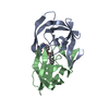
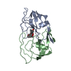
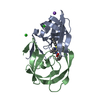
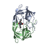
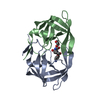
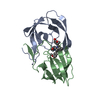
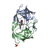
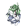
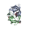
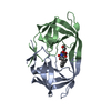
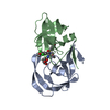
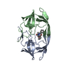
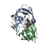
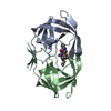
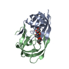
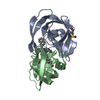
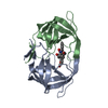
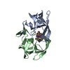
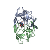
- PDB-1upj: HIV-1 PROTEASE COMPLEX WITH U095438 [3-[1-(4-BROMOPHENYL) ISOBUTY... -
+ Open data

- Basic information
| Entry | Database: PDB / ID: 1upj | ||||||
|---|---|---|---|---|---|---|---|
| Title | HIV-1 PROTEASE COMPLEX WITH U095438 [3-[1-(4-BROMOPHENYL) ISOBUTYL]-4-HYDROXYCOUMARIN | ||||||
 Components Components | HIV-1 PROTEASE | ||||||
 Keywords Keywords | HYDROLASE (ACID PROTEASE) | ||||||
| Function / homology |  Function and homology information Function and homology informationHIV-1 retropepsin / symbiont-mediated activation of host apoptosis / retroviral ribonuclease H / exoribonuclease H / exoribonuclease H activity / DNA integration / viral genome integration into host DNA / establishment of integrated proviral latency / RNA-directed DNA polymerase / RNA stem-loop binding ...HIV-1 retropepsin / symbiont-mediated activation of host apoptosis / retroviral ribonuclease H / exoribonuclease H / exoribonuclease H activity / DNA integration / viral genome integration into host DNA / establishment of integrated proviral latency / RNA-directed DNA polymerase / RNA stem-loop binding / viral penetration into host nucleus / host multivesicular body / RNA-directed DNA polymerase activity / RNA-DNA hybrid ribonuclease activity / Transferases; Transferring phosphorus-containing groups; Nucleotidyltransferases / host cell / viral nucleocapsid / DNA recombination / DNA-directed DNA polymerase / aspartic-type endopeptidase activity / Hydrolases; Acting on ester bonds / DNA-directed DNA polymerase activity / symbiont-mediated suppression of host gene expression / viral translational frameshifting / symbiont entry into host cell / lipid binding / host cell nucleus / host cell plasma membrane / virion membrane / structural molecule activity / proteolysis / DNA binding / zinc ion binding Similarity search - Function | ||||||
| Biological species |   Human immunodeficiency virus 1 Human immunodeficiency virus 1 | ||||||
| Method |  X-RAY DIFFRACTION / MOLECULAR REPLACEMENT / Resolution: 2.22 Å X-RAY DIFFRACTION / MOLECULAR REPLACEMENT / Resolution: 2.22 Å | ||||||
 Authors Authors | Watenpaugh, K.D. / Mulichak, A.M. / Janakiraman, M.N. | ||||||
 Citation Citation | Journal: J.Med.Chem. / Year: 1995 Title: Structure-based design of novel HIV protease inhibitors: carboxamide-containing 4-hydroxycoumarins and 4-hydroxy-2-pyrones as potent nonpeptidic inhibitors. Authors: Thaisrivongs, S. / Watenpaugh, K.D. / Howe, W.J. / Tomich, P.K. / Dolak, L.A. / Chong, K.T. / Tomich, C.C. / Tomasselli, A.G. / Turner, S.R. / Strohbach, J.W. / Mulichak, A.M. / Janakiraman, ...Authors: Thaisrivongs, S. / Watenpaugh, K.D. / Howe, W.J. / Tomich, P.K. / Dolak, L.A. / Chong, K.T. / Tomich, C.C. / Tomasselli, A.G. / Turner, S.R. / Strohbach, J.W. / Mulichak, A.M. / Janakiraman, M.N. / Moon, J.B. / Lynn, J.C. / Horng, M.M. / Hinshaw, R.R. / Curry, K.A. / Rothroc, D.J. | ||||||
| History |
|
- Structure visualization
| Structure viewer | Molecule: MolmilJmol/JSmol |
|---|
- Downloads & links
-Download
| PDBx/mmCIF format | 1upj.cif.gz | 35.4 KB | Display | PDBx/mmCIF format |
|---|---|---|---|---|
| PDB format | pdb1upj.ent.gz | 23 KB | Display | PDB format |
| PDBx/mmJSON format | 1upj.json.gz | Tree view | PDBx/mmJSON format | |
| Others |  Other downloads Other downloads |
-Validation report
| Arichive directory | https://data.pdbj.org/pub/pdb/validation_reports/up/1upjftp://data.pdbj.org/pub/pdb/validation_reports/up/1upj | HTTPS FTP |
|---|
-Related structure data


-Links
 PDBj
PDBj


- Assembly
| Deposited unit | 
| ||||||||||||
|---|---|---|---|---|---|---|---|---|---|---|---|---|---|
| 1 | 
| ||||||||||||
| Unit cell |
| ||||||||||||
| Components on special symmetry positions |
|

-Components
| #1: Protein | Mass: 10803.756 Da / Num. of mol.: 1 Source method: isolated from a genetically manipulated source Source: (gene. exp.) Human immunodeficiency virus 1 / Genus: Lentivirus / Strain: BH5 / Production host:  |
|---|---|
| #2: Chemical | ChemComp-U01 /   Mass: 373.240 Da / Num. of mol.: 1 / Source method: obtained synthetically / Formula: C19H17BrO3 Mass: 373.240 Da / Num. of mol.: 1 / Source method: obtained synthetically / Formula: C19H17BrO3 |
| #3: Water | ChemComp-HOH /  Mass: 18.015 Da / Num. of mol.: 93 / Source method: isolated from a natural source / Formula: H2O Mass: 18.015 Da / Num. of mol.: 93 / Source method: isolated from a natural source / Formula: H2O |
| Nonpolymer details | REGARDING RESIDUE U01 [THE INHIBITOR]: THE ATOMS OA2 AND OA3 ARE, RESPECTIVELY, THE CARBONYL OXYGEN ...REGARDING RESIDUE U01 [THE INHIBITOR]: THE ATOMS OA2 AND OA3 ARE, RESPECTIVE |
-Experimental details
-Experiment
| Experiment | Method: X-RAY DIFFRACTION / Number of used crystals: 1 |
|---|
- Sample preparation
| Crystal | Density Matthews: 2.26 Å3/Da / Density % sol: 45.63 % | ||||||||||||||||||
|---|---|---|---|---|---|---|---|---|---|---|---|---|---|---|---|---|---|---|---|
| Crystal grow | pH: 5.4 / Details: pH 5.4 | ||||||||||||||||||
| Crystal grow | *PLUS Method: vapor diffusion, hanging drop | ||||||||||||||||||
| Components of the solutions | *PLUS
|
-Data collection
| Diffraction | Mean temperature: 298 K |
|---|---|
| Diffraction source | Source: ROTATING ANODE / Type: SIEMENS / Wavelength: 1.5418 |
| Detector | Type: SIEMENS / Detector: AREA DETECTOR / Date: Jan 6, 1992 |
| Radiation | Monochromator: GRAPHITE(002) / Monochromatic (M) / Laue (L): M / Scattering type: x-ray |
| Radiation wavelength | Wavelength: 1.5418 Å / Relative weight: 1 |
| Reflection | Resolution: 2.22→10 Å / Num. obs: 4806 / % possible obs: 89.5 % / Observed criterion σ(I): 0 / Redundancy: 6 % / Rmerge(I) obs: 0.097 / Net I/σ(I): 10.2 |
| Reflection shell | Resolution: 2.22→2.36 Å / Redundancy: 3 % / Rmerge(I) obs: 0.405 / Mean I/σ(I) obs: 1.8 / % possible all: 46 |
| Reflection | *PLUS Num. measured all: 31369 |
| Reflection shell | *PLUS % possible obs: 46 % |
- Processing
| Software |
| ||||||||||||||||||||||||||||||||||||||||||||||||||||||||||||
|---|---|---|---|---|---|---|---|---|---|---|---|---|---|---|---|---|---|---|---|---|---|---|---|---|---|---|---|---|---|---|---|---|---|---|---|---|---|---|---|---|---|---|---|---|---|---|---|---|---|---|---|---|---|---|---|---|---|---|---|---|---|
| Refinement | Method to determine structure: MOLECULAR REPLACEMENT Starting model: EARLIER STRUCTURE Resolution: 2.22→10 Å / σ(F): 2 Details: AS ONLY ONE MOLECULE OF THE INHIBITOR CAN INHIBIT THE DIMERIC PROTEASE BY BINDING TO ITS ACTIVE SITE, THE ATOMS BELONGING TO THE INHIBITOR MOLECULE ARE [TO BE] GIVEN AN OCCUPANCY FACTOR OF 0. ...Details: AS ONLY ONE MOLECULE OF THE INHIBITOR CAN INHIBIT THE DIMERIC PROTEASE BY BINDING TO ITS ACTIVE SITE, THE ATOMS BELONGING TO THE INHIBITOR MOLECULE ARE [TO BE] GIVEN AN OCCUPANCY FACTOR OF 0.5 EACH IN THE SPACE GROUP P6(1)22. NO ALTERNATE CONFORMATIONS ARE OBSERVED FOR THE INHIBITOR. NO ELECTRON DENSITY WAS OBSERVED BEYOND THE POSITION OF THE BETA-CARBON FOR GLU 34, ARG 41, PHE 53, AND GLN 61, THE DELTA-CARBON OF LYS 45, AND LYS 55, AND THE EPSILON-CARBON OF RESIDUE ARG 57. THEY WERE PRESENT DURING REFINEMENT, HOWEVER. IN ADDITION, ATOMS WITH B-FACTORS GREATER THAN 70.0 A**2 MAY BE CONSIDERED TO BE DISORDERED OR NOT SEEN IN THE ELECTRON DENSITY MAPS. SIDE CHAIN ATOMS OF THE THE PHENYL GROUP OF RESIDUE PHE 53 HAVE BEEN MODELED IN ONLY ONE OF THE TWO ALTERNATE CONFORMATIONS AND ASSIGNED AN OCCUPANCY OF 0.5. NO ATOMS IN THE FORM OF DUMMY WATER MOLECULES ARE INTRODUCED AT THE SITE CORRESPONDING TO THE ALTERNATE CONFORMATION. SEE REMARK 6 ALSO. THE BROMINE ATOM OF THE INHIBITOR WAS TREATED AS "S" WITH AN OCCUPANCY OF UNITY DURING CRYSTALLOGRAPHIC REFINEMENTS.
| ||||||||||||||||||||||||||||||||||||||||||||||||||||||||||||
| Refinement step | Cycle: LAST / Resolution: 2.22→10 Å
| ||||||||||||||||||||||||||||||||||||||||||||||||||||||||||||
| Refine LS restraints |
| ||||||||||||||||||||||||||||||||||||||||||||||||||||||||||||
| Software | *PLUS Name: CEDAR / Classification: refinement | ||||||||||||||||||||||||||||||||||||||||||||||||||||||||||||
| Refinement | *PLUS Rfactor obs: 0.178 | ||||||||||||||||||||||||||||||||||||||||||||||||||||||||||||
| Solvent computation | *PLUS | ||||||||||||||||||||||||||||||||||||||||||||||||||||||||||||
| Displacement parameters | *PLUS |
