 Movie
Movie Controller

[English] 日本語
 Yorodumi
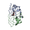
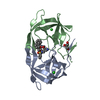
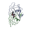
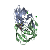
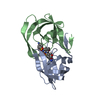
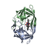
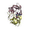
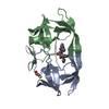
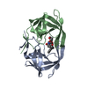
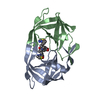
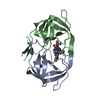
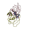
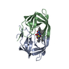
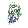
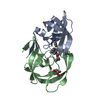
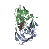
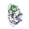
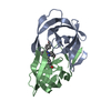
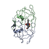
- PDB-2upj: HIV-1 PROTEASE COMPLEX WITH U100313 ([3-[[3-[CYCLOPROPYL [4-HYDRO... -
+ Open data

- Basic information
| Entry | Database: PDB / ID: 2upj | ||||||
|---|---|---|---|---|---|---|---|
| Title | HIV-1 PROTEASE COMPLEX WITH U100313 ([3-[[3-[CYCLOPROPYL [4-HYDROXY-2OXO-6-[1-(PHENYLMETHYL)PROPYL]-2H-PYRAN-3-YL] METHYL]PHENYL]AMINO]-3-OXO-PROPYL]CARBAMIC ACID TERT-BUTYL ESTER) | ||||||
 Components Components | HIV-1 PROTEASE | ||||||
 Keywords Keywords | HYDROLASE (ACID PROTEASE) | ||||||
| Function / homology |  Function and homology information Function and homology informationHIV-1 retropepsin / symbiont-mediated activation of host apoptosis / retroviral ribonuclease H / exoribonuclease H / exoribonuclease H activity / DNA integration / viral genome integration into host DNA / establishment of integrated proviral latency / RNA-directed DNA polymerase / RNA stem-loop binding ...HIV-1 retropepsin / symbiont-mediated activation of host apoptosis / retroviral ribonuclease H / exoribonuclease H / exoribonuclease H activity / DNA integration / viral genome integration into host DNA / establishment of integrated proviral latency / RNA-directed DNA polymerase / RNA stem-loop binding / viral penetration into host nucleus / host multivesicular body / RNA-directed DNA polymerase activity / RNA-DNA hybrid ribonuclease activity / Transferases; Transferring phosphorus-containing groups; Nucleotidyltransferases / host cell / viral nucleocapsid / DNA recombination / DNA-directed DNA polymerase / aspartic-type endopeptidase activity / Hydrolases; Acting on ester bonds / DNA-directed DNA polymerase activity / symbiont-mediated suppression of host gene expression / viral translational frameshifting / symbiont entry into host cell / lipid binding / host cell nucleus / host cell plasma membrane / virion membrane / structural molecule activity / proteolysis / DNA binding / zinc ion binding Similarity search - Function | ||||||
| Biological species |   Human immunodeficiency virus 1 Human immunodeficiency virus 1 | ||||||
| Method |  X-RAY DIFFRACTION / MOLECULAR REPLACEMENT / Resolution: 3 Å X-RAY DIFFRACTION / MOLECULAR REPLACEMENT / Resolution: 3 Å | ||||||
 Authors Authors | Watenpaugh, K.D. / Mulichak, A.M. / Janakiraman, M.N. | ||||||
 Citation Citation | Journal: J.Med.Chem. / Year: 1995 Title: Structure-based design of novel HIV protease inhibitors: carboxamide-containing 4-hydroxycoumarins and 4-hydroxy-2-pyrones as potent nonpeptidic inhibitors. Authors: Thaisrivongs, S. / Watenpaugh, K.D. / Howe, W.J. / Tomich, P.K. / Dolak, L.A. / Chong, K.-T. / Tomich, C.-S.C. / Tomasselli, A.G. / Turner, S.R. / Strohbach, J.W. / Mulichak, A.M. / ...Authors: Thaisrivongs, S. / Watenpaugh, K.D. / Howe, W.J. / Tomich, P.K. / Dolak, L.A. / Chong, K.-T. / Tomich, C.-S.C. / Tomasselli, A.G. / Turner, S.R. / Strohbach, J.W. / Mulichak, A.M. / Janakiraman, M.N. / Moon, J.B. / Lynn, J.C. / Horng, M.-M. / Hinshaw, R.R. / Curry, K.A. / Rothrock, D.J. | ||||||
| History |
|
- Structure visualization
| Structure viewer | Molecule: MolmilJmol/JSmol |
|---|
- Downloads & links
-Download
| PDBx/mmCIF format | 2upj.cif.gz | 51.4 KB | Display | PDBx/mmCIF format |
|---|---|---|---|---|
| PDB format | pdb2upj.ent.gz | 36.4 KB | Display | PDB format |
| PDBx/mmJSON format | 2upj.json.gz | Tree view | PDBx/mmJSON format | |
| Others |  Other downloads Other downloads |
-Validation report
| Arichive directory | https://data.pdbj.org/pub/pdb/validation_reports/up/2upjftp://data.pdbj.org/pub/pdb/validation_reports/up/2upj | HTTPS FTP |
|---|
-Related structure data


-Links
 PDBj
PDBj


- Assembly
| Deposited unit | 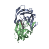
| ||||||||
|---|---|---|---|---|---|---|---|---|---|
| 1 |
| ||||||||
| Unit cell |
|
-Components
| #1: Protein | Mass: 10803.756 Da / Num. of mol.: 2 Source method: isolated from a genetically manipulated source Source: (gene. exp.) Human immunodeficiency virus 1 / Genus: Lentivirus / Strain: BH5 / Production host:  #2: Chemical | ChemComp-U02 / [ |   Mass: 558.665 Da / Num. of mol.: 1 / Source method: obtained synthetically / Formula: C33H38N2O6 Mass: 558.665 Da / Num. of mol.: 1 / Source method: obtained synthetically / Formula: C33H38N2O6#3: Water | ChemComp-HOH / |  Mass: 18.015 Da / Num. of mol.: 56 / Source method: isolated from a natural source / Formula: H2O Mass: 18.015 Da / Num. of mol.: 56 / Source method: isolated from a natural source / Formula: H2ONonpolymer details | THE INHIBITOR MOLECULE BINDS IN ONE ONE ORIENTATION. HOWEVER, THE CARBOXAMIDE SIDE CHAIN WITH ITS ...THE INHIBITOR MOLECULE BINDS IN ONE ONE ORIENTATIO | |
|---|
-Experimental details
-Experiment
| Experiment | Method: X-RAY DIFFRACTION / Number of used crystals: 1 |
|---|
- Sample preparation
| Crystal | Density Matthews: 2.76 Å3/Da / Density % sol: 55.44 % | ||||||||||||||||||
|---|---|---|---|---|---|---|---|---|---|---|---|---|---|---|---|---|---|---|---|
| Crystal grow | pH: 5.2 / Details: pH 5.2 | ||||||||||||||||||
| Crystal grow | *PLUS Method: vapor diffusion, hanging drop | ||||||||||||||||||
| Components of the solutions | *PLUS
|
-Data collection
| Diffraction | Mean temperature: 298 K |
|---|---|
| Diffraction source | Source: ROTATING ANODE / Type: SIEMENS / Wavelength: 1.5418 |
| Detector | Type: SIEMENS / Detector: AREA DETECTOR / Date: Nov 5, 1993 |
| Radiation | Monochromator: GRAPHITE(002) / Monochromatic (M) / Laue (L): M / Scattering type: x-ray |
| Radiation wavelength | Wavelength: 1.5418 Å / Relative weight: 1 |
| Reflection | Resolution: 3→10 Å / Num. obs: 5144 / % possible obs: 100 % / Observed criterion σ(I): 0 / Redundancy: 6 % / Rmerge(I) obs: 0.149 / Net I/σ(I): 6 |
| Reflection shell | Resolution: 3→3.2 Å / Redundancy: 5 % / Rmerge(I) obs: 0.344 / Mean I/σ(I) obs: 3.8 / % possible all: 100 |
| Reflection | *PLUS Num. measured all: 30558 |
- Processing
| Software |
| ||||||||||||||||||||||||||||||||||||||||||||||||||||||||||||
|---|---|---|---|---|---|---|---|---|---|---|---|---|---|---|---|---|---|---|---|---|---|---|---|---|---|---|---|---|---|---|---|---|---|---|---|---|---|---|---|---|---|---|---|---|---|---|---|---|---|---|---|---|---|---|---|---|---|---|---|---|---|
| Refinement | Method to determine structure: MOLECULAR REPLACEMENT Starting model: EARLIER STRUCTURE Resolution: 3→10 Å / σ(F): 2 Details: NO ELECTRON DENSITY WAS OBSERVED BEYOND THE POSITION OF THE BETA-CARBON FOR GLU A 34, ARG A 41, GLN A 61, AND GLN B 18, THE DELTA-CARBON OF LYS A 14, LYS A 43, AND LYS B 55. THEY WERE ...Details: NO ELECTRON DENSITY WAS OBSERVED BEYOND THE POSITION OF THE BETA-CARBON FOR GLU A 34, ARG A 41, GLN A 61, AND GLN B 18, THE DELTA-CARBON OF LYS A 14, LYS A 43, AND LYS B 55. THEY WERE PRESENT DURING REFINEMENT, HOWEVER. IN ADDITION, ATOMS WITH B-FACTORS GREATER THAN 70.0 A**2 MAY BE CONSIDERED TO BE DISORDERED OR NOT SEEN IN THE ELECTRON DENSITY MAPS.
| ||||||||||||||||||||||||||||||||||||||||||||||||||||||||||||
| Refinement step | Cycle: LAST / Resolution: 3→10 Å
| ||||||||||||||||||||||||||||||||||||||||||||||||||||||||||||
| Refine LS restraints |
| ||||||||||||||||||||||||||||||||||||||||||||||||||||||||||||
| Software | *PLUS Name: CEDAR / Classification: refinement | ||||||||||||||||||||||||||||||||||||||||||||||||||||||||||||
| Refinement | *PLUS Rfactor obs: 0.164 | ||||||||||||||||||||||||||||||||||||||||||||||||||||||||||||
| Solvent computation | *PLUS | ||||||||||||||||||||||||||||||||||||||||||||||||||||||||||||
| Displacement parameters | *PLUS |
