 Movie
Movie Controller

[English] 日本語
 Yorodumi
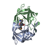
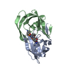
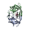
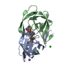
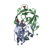
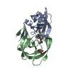
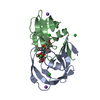
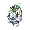
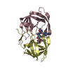
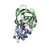
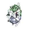
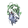
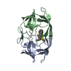
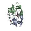
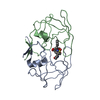
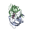
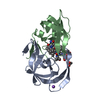
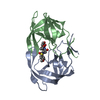
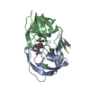
- PDB-6b36: Crystal Structure of HIV Protease complexed with (S)-N-(3-fluoro-... -
+ Open data

- Basic information
| Entry | Database: PDB / ID: 6b36 | ||||||
|---|---|---|---|---|---|---|---|
| Title | Crystal Structure of HIV Protease complexed with (S)-N-(3-fluoro-2-(2-(1-(phenylsulfonyl)piperazin-2-yl)ethyl)phenyl)-3,3-bis(4-fluorophenyl)propanamide | ||||||
 Components Components | HIV-1 Protease | ||||||
 Keywords Keywords | VIRAL PROTEIN/INHIBITOR / HIV / protease / VIRAL PROTEIN / VIRAL PROTEIN-INHIBITOR complex | ||||||
| Function / homology |  Function and homology information Function and homology informationHIV-1 retropepsin / symbiont-mediated activation of host apoptosis / retroviral ribonuclease H / exoribonuclease H / exoribonuclease H activity / DNA integration / viral genome integration into host DNA / establishment of integrated proviral latency / RNA-directed DNA polymerase / RNA stem-loop binding ...HIV-1 retropepsin / symbiont-mediated activation of host apoptosis / retroviral ribonuclease H / exoribonuclease H / exoribonuclease H activity / DNA integration / viral genome integration into host DNA / establishment of integrated proviral latency / RNA-directed DNA polymerase / RNA stem-loop binding / viral penetration into host nucleus / host multivesicular body / RNA-directed DNA polymerase activity / RNA-DNA hybrid ribonuclease activity / Transferases; Transferring phosphorus-containing groups; Nucleotidyltransferases / host cell / viral nucleocapsid / DNA recombination / DNA-directed DNA polymerase / aspartic-type endopeptidase activity / Hydrolases; Acting on ester bonds / DNA-directed DNA polymerase activity / symbiont-mediated suppression of host gene expression / viral translational frameshifting / symbiont entry into host cell / lipid binding / host cell nucleus / host cell plasma membrane / virion membrane / structural molecule activity / proteolysis / DNA binding / zinc ion binding Similarity search - Function | ||||||
| Biological species |   Human immunodeficiency virus 1 Human immunodeficiency virus 1 | ||||||
| Method |  X-RAY DIFFRACTION / SYNCHROTRON / FOURIER SYNTHESIS / Resolution: 1.63 Å X-RAY DIFFRACTION / SYNCHROTRON / FOURIER SYNTHESIS / Resolution: 1.63 Å | ||||||
 Authors Authors | Su, H.P. | ||||||
 Citation Citation | Journal: ACS Med Chem Lett / Year: 2017 Title: Design and Synthesis of Piperazine Sulfonamide Cores Leading to Highly Potent HIV-1 Protease Inhibitors. Authors: Bungard, C.J. / Williams, P.D. / Schulz, J. / Wiscount, C.M. / Holloway, M.K. / Loughran, H.M. / Manikowski, J.J. / Su, H.P. / Bennett, D.J. / Chang, L. / Chu, X.J. / Crespo, A. / Dwyer, M.P. ...Authors: Bungard, C.J. / Williams, P.D. / Schulz, J. / Wiscount, C.M. / Holloway, M.K. / Loughran, H.M. / Manikowski, J.J. / Su, H.P. / Bennett, D.J. / Chang, L. / Chu, X.J. / Crespo, A. / Dwyer, M.P. / Keertikar, K. / Morriello, G.J. / Stamford, A.W. / Waddell, S.T. / Zhong, B. / Hu, B. / Ji, T. / Diamond, T.L. / Bahnck-Teets, C. / Carroll, S.S. / Fay, J.F. / Min, X. / Morris, W. / Ballard, J.E. / Miller, M.D. / McCauley, J.A. | ||||||
| History |
|
- Structure visualization
| Structure viewer | Molecule: MolmilJmol/JSmol |
|---|
- Downloads & links
-Download
| PDBx/mmCIF format | 6b36.cif.gz | 57.4 KB | Display | PDBx/mmCIF format |
|---|---|---|---|---|
| PDB format | pdb6b36.ent.gz | 39.8 KB | Display | PDB format |
| PDBx/mmJSON format | 6b36.json.gz | Tree view | PDBx/mmJSON format | |
| Others |  Other downloads Other downloads |
-Validation report
| Arichive directory | https://data.pdbj.org/pub/pdb/validation_reports/b3/6b36ftp://data.pdbj.org/pub/pdb/validation_reports/b3/6b36 | HTTPS FTP |
|---|
-Related structure data
| Related structure data | 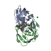 6b38C 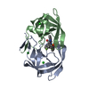 6b3cC 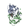 6b3fC 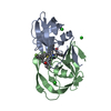 6b3gC 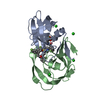 6b3hC  5ivtS S: Starting model for refinement C: citing same article ( |
|---|---|
| Similar structure data |

-Links
 PDBj
PDBj
- Assembly
| Deposited unit | 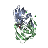
| ||||||||
|---|---|---|---|---|---|---|---|---|---|
| 1 |
| ||||||||
| Unit cell |
|
-Components
| #1: Protein | Mass: 10804.808 Da / Num. of mol.: 2 Source method: isolated from a genetically manipulated source Source: (gene. exp.) Human immunodeficiency virus 1 / Gene: gag-pol / Production host: Human immunodeficiency virus 1 / References: UniProt: P04587, HIV-1 retropepsin#2: Chemical | ChemComp-CL /   Mass: 35.453 Da / Num. of mol.: 4 / Source method: obtained synthetically / Formula: Cl Mass: 35.453 Da / Num. of mol.: 4 / Source method: obtained synthetically / Formula: Cl#3: Chemical | ChemComp-CKD / ( |   Mass: 607.686 Da / Num. of mol.: 1 / Source method: obtained synthetically / Formula: C33H32F3N3O3S Mass: 607.686 Da / Num. of mol.: 1 / Source method: obtained synthetically / Formula: C33H32F3N3O3S#4: Water | ChemComp-HOH / |  Mass: 18.015 Da / Num. of mol.: 147 / Source method: isolated from a natural source / Formula: H2O Mass: 18.015 Da / Num. of mol.: 147 / Source method: isolated from a natural source / Formula: H2O |
|---|
-Experimental details
-Experiment
| Experiment | Method: X-RAY DIFFRACTION / Number of used crystals: 1 |
|---|
- Sample preparation
| Crystal | Density Matthews: 2.66 Å3/Da / Density % sol: 53.74 % |
|---|---|
| Crystal grow | Temperature: 298 K / Method: vapor diffusion, hanging drop / Details: 100 mM sodium acetate, pH 5.0-5.5 300-500mM NaCl |
-Data collection
| Diffraction | Mean temperature: 100 K |
|---|---|
| Diffraction source | Source: SYNCHROTRON / Site: APS  / Beamline: 17-ID / Wavelength: 1 Å / Beamline: 17-ID / Wavelength: 1 Å |
| Detector | Type: DECTRIS PILATUS 6M / Detector: PIXEL / Date: Mar 15, 2017 |
| Radiation | Protocol: SINGLE WAVELENGTH / Monochromatic (M) / Laue (L): M / Scattering type: x-ray |
| Radiation wavelength | Wavelength: 1 Å / Relative weight: 1 |
| Reflection | Resolution: 1.631→19.31 Å / Num. obs: 153989 / % possible obs: 89.3 % / Redundancy: 5.9 % / Biso Wilson estimate: 24.25 Å2 / Net I/σ(I): 19.3 |
- Processing
| Software |
| ||||||||||||||||||||||||||||||||||||||||||||||||||||||||||||||||||||||||||||||||||||||||||||||||||||||||||||||||||
|---|---|---|---|---|---|---|---|---|---|---|---|---|---|---|---|---|---|---|---|---|---|---|---|---|---|---|---|---|---|---|---|---|---|---|---|---|---|---|---|---|---|---|---|---|---|---|---|---|---|---|---|---|---|---|---|---|---|---|---|---|---|---|---|---|---|---|---|---|---|---|---|---|---|---|---|---|---|---|---|---|---|---|---|---|---|---|---|---|---|---|---|---|---|---|---|---|---|---|---|---|---|---|---|---|---|---|---|---|---|---|---|---|---|---|---|
| Refinement | Method to determine structure: FOURIER SYNTHESIS Starting model: 5ivt Resolution: 1.63→19.31 Å / Cor.coef. Fo:Fc: 0.959 / Cor.coef. Fo:Fc free: 0.956 / SU R Cruickshank DPI: 0.092 / Cross valid method: THROUGHOUT / σ(F): 0 / SU R Blow DPI: 0.096 / SU Rfree Blow DPI: 0.09 / SU Rfree Cruickshank DPI: 0.087
| ||||||||||||||||||||||||||||||||||||||||||||||||||||||||||||||||||||||||||||||||||||||||||||||||||||||||||||||||||
| Displacement parameters | Biso mean: 26.43 Å2
| ||||||||||||||||||||||||||||||||||||||||||||||||||||||||||||||||||||||||||||||||||||||||||||||||||||||||||||||||||
| Refine analyze | Luzzati coordinate error obs: 0.21 Å | ||||||||||||||||||||||||||||||||||||||||||||||||||||||||||||||||||||||||||||||||||||||||||||||||||||||||||||||||||
| Refinement step | Cycle: 1 / Resolution: 1.63→19.31 Å
| ||||||||||||||||||||||||||||||||||||||||||||||||||||||||||||||||||||||||||||||||||||||||||||||||||||||||||||||||||
| Refine LS restraints |
| ||||||||||||||||||||||||||||||||||||||||||||||||||||||||||||||||||||||||||||||||||||||||||||||||||||||||||||||||||
| LS refinement shell | Resolution: 1.63→1.7 Å / Total num. of bins used: 13
|
