 Movie
Movie Controller

[English] 日本語
 Yorodumi
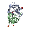
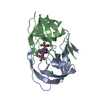
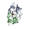
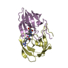
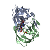
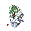
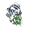
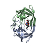
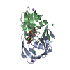
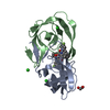
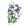
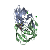
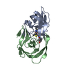
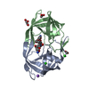
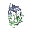
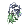
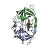
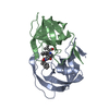
- PDB-3nu6: Crystal Structure of HIV-1 Protease Mutant I54M with Antiviral Dr... -
+ Open data

- Basic information
| Entry | Database: PDB / ID: 3nu6 | ||||||
|---|---|---|---|---|---|---|---|
| Title | Crystal Structure of HIV-1 Protease Mutant I54M with Antiviral Drug Amprenavir | ||||||
 Components Components | Protease | ||||||
 Keywords Keywords | HYDROLASE/HYDROLASE INHIBITOR / enzyme inhibition / aspartic protease / HIV/AIDS / Amprenavir / conformational change / HYDROLASE / HYDROLASE-HYDROLASE INHIBITOR complex | ||||||
| Function / homology |  Function and homology information Function and homology informationHIV-1 retropepsin / symbiont-mediated activation of host apoptosis / retroviral ribonuclease H / exoribonuclease H / exoribonuclease H activity / DNA integration / viral genome integration into host DNA / establishment of integrated proviral latency / RNA-directed DNA polymerase / RNA stem-loop binding ...HIV-1 retropepsin / symbiont-mediated activation of host apoptosis / retroviral ribonuclease H / exoribonuclease H / exoribonuclease H activity / DNA integration / viral genome integration into host DNA / establishment of integrated proviral latency / RNA-directed DNA polymerase / RNA stem-loop binding / viral penetration into host nucleus / host multivesicular body / RNA-directed DNA polymerase activity / RNA-DNA hybrid ribonuclease activity / Transferases; Transferring phosphorus-containing groups; Nucleotidyltransferases / host cell / viral nucleocapsid / DNA recombination / DNA-directed DNA polymerase / aspartic-type endopeptidase activity / Hydrolases; Acting on ester bonds / DNA-directed DNA polymerase activity / symbiont-mediated suppression of host gene expression / viral translational frameshifting / symbiont entry into host cell / lipid binding / host cell nucleus / host cell plasma membrane / virion membrane / structural molecule activity / proteolysis / DNA binding / zinc ion binding Similarity search - Function | ||||||
| Biological species |   Human immunodeficiency virus 1 Human immunodeficiency virus 1 | ||||||
| Method |  X-RAY DIFFRACTION / SYNCHROTRON / AB INITIO / Resolution: 1.16 Å X-RAY DIFFRACTION / SYNCHROTRON / AB INITIO / Resolution: 1.16 Å | ||||||
 Authors Authors | Shen, C.H. / Weber, I.T. | ||||||
 Citation Citation | Journal: Febs J. / Year: 2010 Title: Amprenavir complexes with HIV-1 protease and its drug-resistant mutants altering hydrophobic clusters. Authors: Shen, C.H. / Wang, Y.F. / Kovalevsky, A.Y. / Harrison, R.W. / Weber, I.T. | ||||||
| History |
|
- Structure visualization
| Structure viewer | Molecule: MolmilJmol/JSmol |
|---|
- Downloads & links
-Download
| PDBx/mmCIF format | 3nu6.cif.gz | 107.2 KB | Display | PDBx/mmCIF format |
|---|---|---|---|---|
| PDB format | pdb3nu6.ent.gz | 80.8 KB | Display | PDB format |
| PDBx/mmJSON format | 3nu6.json.gz | Tree view | PDBx/mmJSON format | |
| Others |  Other downloads Other downloads |
-Validation report
| Arichive directory | https://data.pdbj.org/pub/pdb/validation_reports/nu/3nu6ftp://data.pdbj.org/pub/pdb/validation_reports/nu/3nu6 | HTTPS FTP |
|---|
-Related structure data
| Related structure data | 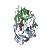 3nu3C 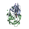 3nu4C  3nu5C 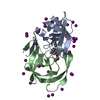 3nu9C 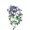 3nujC  3nuoC  2f8gS C: citing same article ( S: Starting model for refinement |
|---|---|
| Similar structure data |
-Links
 PDBj
PDBj


- Assembly
| Deposited unit | 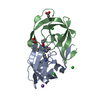
| ||||||||
|---|---|---|---|---|---|---|---|---|---|
| 1 |
| ||||||||
| Unit cell |
| ||||||||
| Components on special symmetry positions |
|
-Components
-Protein , 1 types, 2 molecules AB
| #1: Protein | Mass: 10758.715 Da / Num. of mol.: 2 / Fragment: residues 501-599 / Mutation: Q7K, L33I, I54M, L63I, C67A, C95A Source method: isolated from a genetically manipulated source Source: (gene. exp.) Human immunodeficiency virus 1 / Genus: Lentivirus / Gene: gag, pol / Plasmid: PET11A / Species (production host): Escherichia coli / Production host:  |
|---|
-Non-polymers , 5 types, 239 molecules 


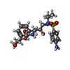

| #2: Chemical |  Mass: 35.453 Da / Num. of mol.: 3 / Source method: obtained synthetically / Formula: Cl Mass: 35.453 Da / Num. of mol.: 3 / Source method: obtained synthetically / Formula: Cl#3: Chemical | ChemComp-NA / |  Mass: 22.990 Da / Num. of mol.: 1 / Source method: obtained synthetically / Formula: Na Mass: 22.990 Da / Num. of mol.: 1 / Source method: obtained synthetically / Formula: Na#4: Chemical |  Mass: 59.044 Da / Num. of mol.: 2 / Source method: obtained synthetically / Formula: C2H3O2 Mass: 59.044 Da / Num. of mol.: 2 / Source method: obtained synthetically / Formula: C2H3O2#5: Chemical | ChemComp-478 / { |  Mass: 505.627 Da / Num. of mol.: 1 / Source method: obtained synthetically / Formula: C25H35N3O6S / Comment: medication*YM Mass: 505.627 Da / Num. of mol.: 1 / Source method: obtained synthetically / Formula: C25H35N3O6S / Comment: medication*YM#6: Water | ChemComp-HOH / | Mass: 18.015 Da / Num. of mol.: 232 / Source method: isolated from a natural source / Formula: H2O |
|---|
-Experimental details
-Experiment
| Experiment | Method: X-RAY DIFFRACTION / Number of used crystals: 1 |
|---|
- Sample preparation
| Crystal | Density Matthews: 2.67 Å3/Da / Density % sol: 54.01 % |
|---|---|
| Crystal grow | Temperature: 298 K / Method: vapor diffusion, hanging drop / pH: 4.6 Details: Crystal was grown by the hanging-drop vapor-diffusion method at room temperature, from a 2.2 mg/ml protein solution at pH 4.6 with 0.1M sodium acetate, 0.67M sodium chloride. The inhibitor ...Details: Crystal was grown by the hanging-drop vapor-diffusion method at room temperature, from a 2.2 mg/ml protein solution at pH 4.6 with 0.1M sodium acetate, 0.67M sodium chloride. The inhibitor was mixed with protease in a ratio 5:1 , VAPOR DIFFUSION, HANGING DROP, temperature 298K |
-Data collection
| Diffraction | Mean temperature: 100 K |
|---|---|
| Diffraction source | Source: SYNCHROTRON / Site: APS  / Beamline: 22-ID / Wavelength: 1 Å / Beamline: 22-ID / Wavelength: 1 Å |
| Detector | Type: MARMOSAIC 300 mm CCD / Detector: CCD / Date: Jul 24, 2007 |
| Radiation | Protocol: SINGLE WAVELENGTH / Monochromatic (M) / Laue (L): M / Scattering type: x-ray |
| Radiation wavelength | Wavelength: 1 Å / Relative weight: 1 |
| Reflection | Resolution: 1.16→50 Å / % possible obs: 91.8 % / Observed criterion σ(F): 0 / Observed criterion σ(I): 0 / Redundancy: 5 % / Biso Wilson estimate: 10.6 Å2 / Rmerge(I) obs: 0.072 |
| Reflection shell | Resolution: 1.16→1.2 Å / Redundancy: 2.1 % / Rmerge(I) obs: 0.357 / Mean I/σ(I) obs: 2 / % possible all: 58.9 |
- Processing
| Software |
| |||||||||||||||||||||||||||||||||
|---|---|---|---|---|---|---|---|---|---|---|---|---|---|---|---|---|---|---|---|---|---|---|---|---|---|---|---|---|---|---|---|---|---|---|
| Refinement | Method to determine structure: AB INITIO Starting model: 2F8G Resolution: 1.16→10 Å / Num. parameters: 16993 / Num. restraintsaints: 21671 / Cross valid method: FREE R / σ(F): 0 / Stereochemistry target values: ENGH AND HUBER / Details: conjugate gradient minimization
| |||||||||||||||||||||||||||||||||
| Refine analyze | Num. disordered residues: 11 / Occupancy sum hydrogen: 1648.37 / Occupancy sum non hydrogen: 1765.77 | |||||||||||||||||||||||||||||||||
| Refinement step | Cycle: LAST / Resolution: 1.16→10 Å
| |||||||||||||||||||||||||||||||||
| Refine LS restraints |
|
