 Movie
Movie Controller

[English] 日本語
 Yorodumi
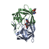
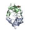
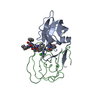
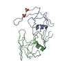
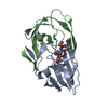
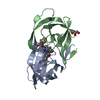
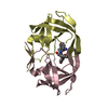
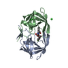
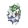
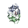
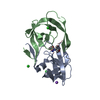
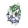
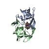
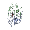
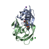
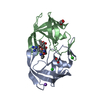
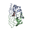
- PDB-4j54: Crystal Structure of Multidrug Resistant HIV-1 Protease Clinical ... -
+ Open data

- Basic information
| Entry | Database: PDB / ID: 4j54 | ||||||
|---|---|---|---|---|---|---|---|
| Title | Crystal Structure of Multidrug Resistant HIV-1 Protease Clinical isolate PR20 with the potent antiviral inhibitor GRL-0519A | ||||||
 Components Components | Protease | ||||||
 Keywords Keywords | HYDROLASE/HYDROLASE INHIBITOR / multidrug resistant HIV-1 protease clinical isolate PR20 / clinical inhibitor Amprenavir / potent antiviral inhibitor GRL-0519A / potent antiviral inhibitor GRL-02031 / HYDROLASE-HYDROLASE INHIBITOR complex | ||||||
| Function / homology |  Function and homology information Function and homology informationHIV-1 retropepsin / symbiont-mediated activation of host apoptosis / retroviral ribonuclease H / exoribonuclease H / exoribonuclease H activity / DNA integration / viral genome integration into host DNA / establishment of integrated proviral latency / RNA-directed DNA polymerase / RNA stem-loop binding ...HIV-1 retropepsin / symbiont-mediated activation of host apoptosis / retroviral ribonuclease H / exoribonuclease H / exoribonuclease H activity / DNA integration / viral genome integration into host DNA / establishment of integrated proviral latency / RNA-directed DNA polymerase / RNA stem-loop binding / viral penetration into host nucleus / host multivesicular body / RNA-directed DNA polymerase activity / RNA-DNA hybrid ribonuclease activity / Transferases; Transferring phosphorus-containing groups; Nucleotidyltransferases / host cell / viral nucleocapsid / DNA recombination / DNA-directed DNA polymerase / aspartic-type endopeptidase activity / Hydrolases; Acting on ester bonds / DNA-directed DNA polymerase activity / symbiont-mediated suppression of host gene expression / viral translational frameshifting / symbiont entry into host cell / lipid binding / host cell nucleus / host cell plasma membrane / virion membrane / structural molecule activity / proteolysis / DNA binding / zinc ion binding Similarity search - Function | ||||||
| Biological species |   Human immunodeficiency virus 1 Human immunodeficiency virus 1 | ||||||
| Method |  X-RAY DIFFRACTION / SYNCHROTRON / MOLECULAR REPLACEMENT / Resolution: 1.55 Å X-RAY DIFFRACTION / SYNCHROTRON / MOLECULAR REPLACEMENT / Resolution: 1.55 Å | ||||||
 Authors Authors | Wang, Y.-F. / Agniswamy, J. / Weber, I.T. | ||||||
 Citation Citation | Journal: J.Med.Chem. / Year: 2013 Title: Extreme Multidrug Resistant HIV-1 Protease with 20 Mutations Is Resistant to Novel Protease Inhibitors with P1'-Pyrrolidinone or P2-Tris-tetrahydrofuran. Authors: Agniswamy, J. / Shen, C.H. / Wang, Y.F. / Ghosh, A.K. / Rao, K.V. / Xu, C.X. / Sayer, J.M. / Louis, J.M. / Weber, I.T. | ||||||
| History |
|
- Structure visualization
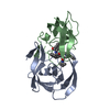
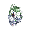
| Structure viewer | Molecule: MolmilJmol/JSmol |
|---|
- Downloads & links
-Download
| PDBx/mmCIF format | 4j54.cif.gz | 58.2 KB | Display | PDBx/mmCIF format |
|---|---|---|---|---|
| PDB format | pdb4j54.ent.gz | 40.5 KB | Display | PDB format |
| PDBx/mmJSON format | 4j54.json.gz | Tree view | PDBx/mmJSON format | |
| Others |  Other downloads Other downloads |
-Validation report
| Arichive directory | https://data.pdbj.org/pub/pdb/validation_reports/j5/4j54ftp://data.pdbj.org/pub/pdb/validation_reports/j5/4j54 | HTTPS FTP |
|---|
-Related structure data
| Related structure data | 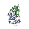 4j55C 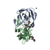 4j5jC 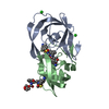 2hs2S C: citing same article ( S: Starting model for refinement |
|---|---|
| Similar structure data |


-Links
 PDBj
PDBj


- Assembly
| Deposited unit | 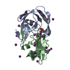
| ||||||||
|---|---|---|---|---|---|---|---|---|---|
| 1 |
| ||||||||
| Unit cell |
|
-Components
| #1: Protein | Mass: 10766.542 Da / Num. of mol.: 2 / Fragment: UNP residues 501-599 Mutation: Q7K,L10F,I13V,I15V,D30N,V32I,L33F,E35D,M36I,S37N,I47V,I54L,Q58E,I62V,L63P,A71V,I84V,N88D,L89T,L90M Source method: isolated from a genetically manipulated source Source: (gene. exp.) Human immunodeficiency virus 1 / Gene: pol / Plasmid: pET11a / Production host:  #2: Chemical | 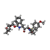  Mass: 604.712 Da / Num. of mol.: 2 / Source method: obtained synthetically / Formula: C30H40N2O9S Mass: 604.712 Da / Num. of mol.: 2 / Source method: obtained synthetically / Formula: C30H40N2O9S#3: Chemical | ChemComp-IOD /   Mass: 126.904 Da / Num. of mol.: 22 / Source method: obtained synthetically / Formula: I Mass: 126.904 Da / Num. of mol.: 22 / Source method: obtained synthetically / Formula: I#4: Chemical | ChemComp-ACT / |   Mass: 59.044 Da / Num. of mol.: 1 / Source method: obtained synthetically / Formula: C2H3O2 Mass: 59.044 Da / Num. of mol.: 1 / Source method: obtained synthetically / Formula: C2H3O2#5: Water | ChemComp-HOH / |  Mass: 18.015 Da / Num. of mol.: 88 / Source method: isolated from a natural source / Formula: H2O Mass: 18.015 Da / Num. of mol.: 88 / Source method: isolated from a natural source / Formula: H2O |
|---|
-Experimental details
-Experiment
| Experiment | Method: X-RAY DIFFRACTION / Number of used crystals: 1 |
|---|
- Sample preparation
| Crystal | Density Matthews: 2.03 Å3/Da / Density % sol: 39.28 % |
|---|---|
| Crystal grow | Temperature: 298 K / Method: vapor diffusion, hanging drop / pH: 4.8 Details: 0.25 M potassium iodide, 0.1 M sodium acetate, pH 4.8, VAPOR DIFFUSION, HANGING DROP, temperature 298K |
-Data collection
| Diffraction | Mean temperature: 100 K |
|---|---|
| Diffraction source | Source: SYNCHROTRON / Site: APS  / Beamline: 22-ID / Wavelength: 0.8 Å / Beamline: 22-ID / Wavelength: 0.8 Å |
| Detector | Type: MARMOSAIC 300 mm CCD / Detector: CCD / Date: Aug 4, 2010 |
| Radiation | Monochromator: Rosenbaum-Rock double-crystal Si(111) / Protocol: SINGLE WAVELENGTH / Monochromatic (M) / Laue (L): M / Scattering type: x-ray |
| Radiation wavelength | Wavelength: 0.8 Å / Relative weight: 1 |
| Reflection | Resolution: 1.55→50 Å / Num. all: 24299 / Num. obs: 24299 / % possible obs: 92.5 % / Redundancy: 6.1 % / Rmerge(I) obs: 0.069 / Net I/σ(I): 22.6 |
| Reflection shell | Resolution: 1.55→1.61 Å / Redundancy: 1.8 % / Rmerge(I) obs: 0.265 / Mean I/σ(I) obs: 2.5 / % possible all: 50.2 |
- Processing
| Software |
| |||||||||||||||||||||||||||||||||
|---|---|---|---|---|---|---|---|---|---|---|---|---|---|---|---|---|---|---|---|---|---|---|---|---|---|---|---|---|---|---|---|---|---|---|
| Refinement | Method to determine structure: MOLECULAR REPLACEMENT Starting model: PDB ENTRY 2HS2 Resolution: 1.55→10 Å / Num. parameters: 6784 / Num. restraintsaints: 6513 / Cross valid method: FREE R / σ(F): 0 / Stereochemistry target values: ENGH AND HUBER
| |||||||||||||||||||||||||||||||||
| Solvent computation | Solvent model: MOEWS & KRETSINGER, J.MOL.BIOL.91(1973)201-228 | |||||||||||||||||||||||||||||||||
| Displacement parameters | Biso mean: 19.8692 Å2 | |||||||||||||||||||||||||||||||||
| Refine analyze | Num. disordered residues: 3 / Occupancy sum hydrogen: 0 / Occupancy sum non hydrogen: 1653.65 | |||||||||||||||||||||||||||||||||
| Refinement step | Cycle: LAST / Resolution: 1.55→10 Å
| |||||||||||||||||||||||||||||||||
| Refine LS restraints |
|
