 Movie
Movie Controller

[English] 日本語
 Yorodumi
- PDB-6mei: Crystal structure of broadly neutralizing antibody HEPC3 in compl... -
+ Open data

- Basic information
| Entry | Database: PDB / ID: 6mei | ||||||||||||
|---|---|---|---|---|---|---|---|---|---|---|---|---|---|
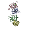
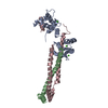
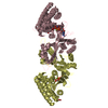
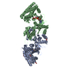
| Title | Crystal structure of broadly neutralizing antibody HEPC3 in complex with Hepatitis C virus envelope glycoprotein E2 ectodomain | ||||||||||||
 Components Components |
| ||||||||||||
 Keywords Keywords | IMMUNE SYSTEM / HCV glycoprotein / broadly neutralizing antibodies | ||||||||||||
| Function / homology |  Function and homology information Function and homology informationhost cell mitochondrial membrane / host cell lipid droplet / viral nucleocapsid / clathrin-dependent endocytosis of virus by host cell / symbiont-mediated suppression of host innate immune response / host cell endoplasmic reticulum membrane / ribonucleoprotein complex / fusion of virus membrane with host endosome membrane / viral envelope / virion attachment to host cell ...host cell mitochondrial membrane / host cell lipid droplet / viral nucleocapsid / clathrin-dependent endocytosis of virus by host cell / symbiont-mediated suppression of host innate immune response / host cell endoplasmic reticulum membrane / ribonucleoprotein complex / fusion of virus membrane with host endosome membrane / viral envelope / virion attachment to host cell / host cell nucleus / virion membrane / structural molecule activity / RNA binding Similarity search - Function | ||||||||||||
| Biological species |  Hepacivirus C Hepacivirus C Homo sapiens (human) Homo sapiens (human) | ||||||||||||
| Method |  X-RAY DIFFRACTION / SYNCHROTRON / Resolution: 2.9 Å X-RAY DIFFRACTION / SYNCHROTRON / Resolution: 2.9 Å | ||||||||||||
 Authors Authors | Flyak, A.I. / Bjorkman, P.J. | ||||||||||||
| Funding support |  United States, 1items United States, 1items
| ||||||||||||
 Citation Citation | Journal: Cell Host Microbe / Year: 2018 Title: HCV Broadly Neutralizing Antibodies Use a CDRH3 Disulfide Motif to Recognize an E2 Glycoprotein Site that Can Be Targeted for Vaccine Design. Authors: Flyak, A.I. / Ruiz, S. / Colbert, M.D. / Luong, T. / Crowe Jr., J.E. / Bailey, J.R. / Bjorkman, P.J. | ||||||||||||
| History |
|
- Structure visualization
| Structure viewer | Molecule: MolmilJmol/JSmol |
|---|
- Downloads & links
-Download
| PDBx/mmCIF format | 6mei.cif.gz | 158.2 KB | Display | PDBx/mmCIF format |
|---|---|---|---|---|
| PDB format | pdb6mei.ent.gz | 120.1 KB | Display | PDB format |
| PDBx/mmJSON format | 6mei.json.gz | Tree view | PDBx/mmJSON format | |
| Others |  Other downloads Other downloads |
-Validation report
| Arichive directory | https://data.pdbj.org/pub/pdb/validation_reports/me/6meiftp://data.pdbj.org/pub/pdb/validation_reports/me/6mei | HTTPS FTP |
|---|
-Related structure data
| Related structure data | 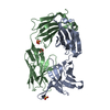 6medC 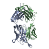 6meeC 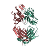 6mefC 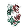 6megC  6mehC  6mejC  6mekC C: citing same article ( |
|---|---|
| Similar structure data |


-Links
 PDBj
PDBj


- Assembly
| Deposited unit | 
| ||||||||
|---|---|---|---|---|---|---|---|---|---|
| 1 |
| ||||||||
| Unit cell |
|
-Components
-Protein , 1 types, 1 molecules C
| #1: Protein | Mass: 29945.557 Da / Num. of mol.: 1 Source method: isolated from a genetically manipulated source Source: (gene. exp.) Hepacivirus C / Plasmid: pCMV / Cell line (production host): HEK293-6E / Production host: Homo sapiens (human) / References: UniProt: A0A2P0NE26 |
|---|
-Antibody , 2 types, 2 molecules HL
| #2: Antibody | Mass: 25759.807 Da / Num. of mol.: 1 Source method: isolated from a genetically manipulated source Source: (gene. exp.) Homo sapiens (human) / Plasmid: pTT5 / Cell line (production host): HEK293-6E / Production host: Homo sapiens (human) |
|---|---|
| #3: Antibody | Mass: 23384.883 Da / Num. of mol.: 1 Source method: isolated from a genetically manipulated source Source: (gene. exp.) Homo sapiens (human) / Plasmid: pTT5 / Cell line (production host): HEK293-6E / Production host: Homo sapiens (human) |
-Sugars , 3 types, 8 molecules 
| #4: Polysaccharide | 2-acetamido-2-deoxy-beta-D-glucopyranose-(1-4)-2-acetamido-2-deoxy-beta-D-glucopyranose Source method: isolated from a genetically manipulated source | ||
|---|---|---|---|
| #5: Polysaccharide | Source method: isolated from a genetically manipulated source #6: Sugar | ChemComp-NAG /  Type: D-saccharide, beta linking / Mass: 221.208 Da / Num. of mol.: 5 Type: D-saccharide, beta linking / Mass: 221.208 Da / Num. of mol.: 5Source method: isolated from a genetically manipulated source Formula: C8H15NO6 |
-Non-polymers , 2 types, 211 molecules 

| #7: Chemical | ChemComp-BR /  Mass: 79.904 Da / Num. of mol.: 7 / Source method: obtained synthetically / Formula: Br Mass: 79.904 Da / Num. of mol.: 7 / Source method: obtained synthetically / Formula: Br#8: Water | ChemComp-HOH / | Mass: 18.015 Da / Num. of mol.: 204 / Source method: isolated from a natural source / Formula: H2O |
|---|
-Details
| Has protein modification | Y |
|---|
-Experimental details
-Experiment
| Experiment | Method: X-RAY DIFFRACTION / Number of used crystals: 1 |
|---|
- Sample preparation
| Crystal | Density Matthews: 2.74 Å3/Da / Density % sol: 55.16 % / Mosaicity: 0.9 ° |
|---|---|
| Crystal grow | Temperature: 298 K / Method: vapor diffusion, hanging drop / Details: 0.2M sodium bromide, 20% PEG 3,350 |
-Data collection
| Diffraction | Mean temperature: 100 K | ||||||||||||||||||||||||||||||
|---|---|---|---|---|---|---|---|---|---|---|---|---|---|---|---|---|---|---|---|---|---|---|---|---|---|---|---|---|---|---|---|
| Diffraction source | Source: SYNCHROTRON / Site: APS / Beamline: 23-ID-D / Wavelength: 1 Å | ||||||||||||||||||||||||||||||
| Detector | Type: DECTRIS PILATUS 6M / Detector: PIXEL / Date: Oct 28, 2017 | ||||||||||||||||||||||||||||||
| Radiation | Protocol: SINGLE WAVELENGTH / Monochromatic (M) / Laue (L): M / Scattering type: x-ray | ||||||||||||||||||||||||||||||
| Radiation wavelength | Wavelength: 1 Å / Relative weight: 1 | ||||||||||||||||||||||||||||||
| Reflection | Resolution: 2.2→87 Å / Num. obs: 44996 / % possible obs: 99.8 % / Redundancy: 5.1 % / Biso Wilson estimate: 37.77 Å2 / CC1/2: 0.885 / Rmerge(I) obs: 0.271 / Rpim(I) all: 0.145 / Rrim(I) all: 0.31 / Net I/σ(I): 3.7 / Num. measured all: 231097 / Scaling rejects: 3510 | ||||||||||||||||||||||||||||||
| Reflection shell | Diffraction-ID: 1
|
- Processing
| Software |
| ||||||||||||||||||||||||||||||||||||||||||||||||||||||||
|---|---|---|---|---|---|---|---|---|---|---|---|---|---|---|---|---|---|---|---|---|---|---|---|---|---|---|---|---|---|---|---|---|---|---|---|---|---|---|---|---|---|---|---|---|---|---|---|---|---|---|---|---|---|---|---|---|---|
| Refinement | Resolution: 2.9→86.999 Å / SU ML: 0.32 / Cross valid method: THROUGHOUT / σ(F): 1.34 / Phase error: 25.9 / Stereochemistry target values: ML
| ||||||||||||||||||||||||||||||||||||||||||||||||||||||||
| Solvent computation | Shrinkage radii: 0.9 Å / VDW probe radii: 1.11 Å / Solvent model: FLAT BULK SOLVENT MODEL | ||||||||||||||||||||||||||||||||||||||||||||||||||||||||
| Displacement parameters | Biso max: 107.24 Å2 / Biso mean: 32.0416 Å2 / Biso min: 7.37 Å2 | ||||||||||||||||||||||||||||||||||||||||||||||||||||||||
| Refinement step | Cycle: final / Resolution: 2.9→86.999 Å
| ||||||||||||||||||||||||||||||||||||||||||||||||||||||||
| LS refinement shell | Refine-ID: X-RAY DIFFRACTION / Rfactor Rfree error: 0 / Total num. of bins used: 7
|
