 Movie
Movie Controller

[English] 日本語
 Yorodumi
- PDB-1j1e: Crystal structure of the 52kDa domain of human cardiac troponin i... -
+ Open data

- Basic information
| Entry | Database: PDB / ID: 1j1e | ||||||
|---|---|---|---|---|---|---|---|
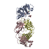
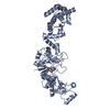
| Title | Crystal structure of the 52kDa domain of human cardiac troponin in the Ca2+ saturated form | ||||||
 Components Components |
| ||||||
 Keywords Keywords | CONTRACTILE PROTEIN / Thin filament / muscle regulation / Ca2+ binding protein / EF-hand / coiled-coil | ||||||
| Function / homology |  Function and homology information Function and homology informationregulation of systemic arterial blood pressure by ischemic conditions / troponin C binding / diaphragm contraction / regulation of muscle filament sliding speed / troponin T binding / cardiac Troponin complex / cardiac myofibril / negative regulation of ATP-dependent activity / troponin complex / regulation of muscle contraction ...regulation of systemic arterial blood pressure by ischemic conditions / troponin C binding / diaphragm contraction / regulation of muscle filament sliding speed / troponin T binding / cardiac Troponin complex / cardiac myofibril / negative regulation of ATP-dependent activity / troponin complex / regulation of muscle contraction / regulation of smooth muscle contraction / transition between fast and slow fiber / positive regulation of ATP-dependent activity / Striated Muscle Contraction / muscle filament sliding / sarcomere organization / regulation of cardiac muscle contraction by calcium ion signaling / response to metal ion / cardiac muscle cell contraction / ventricular cardiac muscle tissue morphogenesis / heart contraction / regulation of heart contraction / tropomyosin binding / troponin I binding / striated muscle thin filament / vasculogenesis / calcium channel inhibitor activity / skeletal muscle contraction / Ion homeostasis / cardiac muscle contraction / sarcomere / response to calcium ion / intracellular calcium ion homeostasis / calcium-dependent protein binding / actin filament binding / heart development / actin binding / protein domain specific binding / calcium ion binding / protein kinase binding / protein homodimerization activity / identical protein binding / cytosol Similarity search - Function | ||||||
| Biological species |  Homo sapiens (human) Homo sapiens (human) | ||||||
| Method |  X-RAY DIFFRACTION / SYNCHROTRON / MOLECULAR REPLACEMENT / Resolution: 3.3 Å X-RAY DIFFRACTION / SYNCHROTRON / MOLECULAR REPLACEMENT / Resolution: 3.3 Å | ||||||
 Authors Authors | Takeda, S. / Yamashita, A. / Maeda, K. / Maeda, Y. | ||||||
 Citation Citation | Journal: Nature / Year: 2003 Title: Structure of the core domain of human cardiac troponin in the Ca2+-saturated form Authors: Takeda, S. / Yamashita, A. / Maeda, K. / Maeda, Y. #1: Journal: Eur.J.Biochem. / Year: 1997Title: Structural and functional domains of the troponin complex revealed by limited digestion Authors: Takeda, S. / Kobayashi, T. / Taniguchi, H. / Hayashi, H. / Maeda, Y. #2: Journal: Proc.Natl.Acad.Sci.USA / Year: 1998Title: Crystal structure of troponin C in complex with troponin I fragment at 2.3-A resolution Authors: Vassylyev, D.G. / Takeda, S. / Wakatsuki, S. / Maeda, K. / Maeda, Y. | ||||||
| History |
|
- Structure visualization
| Structure viewer | Molecule: MolmilJmol/JSmol |
|---|
- Downloads & links
-Download
| PDBx/mmCIF format | 1j1e.cif.gz | 147.4 KB | Display | PDBx/mmCIF format |
|---|---|---|---|---|
| PDB format | pdb1j1e.ent.gz | 116.2 KB | Display | PDB format |
| PDBx/mmJSON format | 1j1e.json.gz | Tree view | PDBx/mmJSON format | |
| Others |  Other downloads Other downloads |
-Validation report
| Arichive directory | https://data.pdbj.org/pub/pdb/validation_reports/j1/1j1eftp://data.pdbj.org/pub/pdb/validation_reports/j1/1j1e | HTTPS FTP |
|---|
-Related structure data
| Related structure data |  1j1dSC S: Starting model for refinement C: citing same article ( |
|---|---|
| Similar structure data |


-Links
 PDBj
PDBj


- Assembly
| Deposited unit | 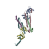
| ||||||||
|---|---|---|---|---|---|---|---|---|---|
| 1 | 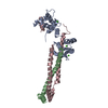
| ||||||||
| 2 | 
| ||||||||
| Unit cell |
| ||||||||
| Details | chain A, B and C, and chain D, E and F are biological heterotrimer assemblies, respectively. |
-Components
| #1: Protein | Mass: 18401.377 Da / Num. of mol.: 2 / Mutation: C35S, C84S Source method: isolated from a genetically manipulated source Source: (gene. exp.) Homo sapiens (human) / Tissue: cardiac muscle / Plasmid: pET3d / Production host:  #2: Protein | Mass: 12842.768 Da / Num. of mol.: 2 / Fragment: CNBR fragment, residues 183-288 Source method: isolated from a genetically manipulated source Source: (gene. exp.) Homo sapiens (human) / Tissue: cardiac muscle / Plasmid: pET3d / Production host: #3: Protein | Mass: 20746.012 Da / Num. of mol.: 2 / Fragment: Residues 31-210 / Mutation: T31M, C80A, C97A Source method: isolated from a genetically manipulated source Source: (gene. exp.) Homo sapiens (human) / Tissue: cardiac muscle / Plasmid: pET3d / Production host: #4: Chemical | ChemComp-CA /   Mass: 40.078 Da / Num. of mol.: 6 / Source method: obtained synthetically / Formula: Ca Mass: 40.078 Da / Num. of mol.: 6 / Source method: obtained synthetically / Formula: Ca |
|---|
-Experimental details
-Experiment
| Experiment | Method: X-RAY DIFFRACTION / Number of used crystals: 1 |
|---|
- Sample preparation
| Crystal | Density Matthews: 3.34 Å3/Da / Density % sol: 62.89 % | |||||||||||||||||||||||||||||||||||||||||||||||||
|---|---|---|---|---|---|---|---|---|---|---|---|---|---|---|---|---|---|---|---|---|---|---|---|---|---|---|---|---|---|---|---|---|---|---|---|---|---|---|---|---|---|---|---|---|---|---|---|---|---|---|
| Crystal grow | Temperature: 293 K / Method: vapor diffusion, hanging drop / pH: 8 Details: PEG3350, lithium chloride, Tris-HCl, calcium chloride, glycerol, pH 8.0, VAPOR DIFFUSION, HANGING DROP, temperature 293K | |||||||||||||||||||||||||||||||||||||||||||||||||
| Crystal grow | *PLUS pH: 8 / Method: vapor diffusion, hanging drop | |||||||||||||||||||||||||||||||||||||||||||||||||
| Components of the solutions | *PLUS
|
-Data collection
| Diffraction | Mean temperature: 90 K |
|---|---|
| Diffraction source | Source: SYNCHROTRON / Site: SPring-8  / Beamline: BL41XU / Wavelength: 1 Å / Beamline: BL41XU / Wavelength: 1 Å |
| Detector | Type: MAR CCD 165 mm / Detector: CCD / Date: Feb 23, 2001 Details: KARKPATRIC-BOETZE TYPE RH-COATED DOUBLE MIRROR (SUPER MIRRORS) |
| Radiation | Monochromator: Si(111) / Protocol: SINGLE WAVELENGTH / Monochromatic (M) / Laue (L): M / Scattering type: x-ray |
| Radiation wavelength | Wavelength: 1 Å / Relative weight: 1 |
| Reflection | Resolution: 3.3→20 Å / Num. all: 16308 / Num. obs: 16101 / % possible obs: 99.3 % / Observed criterion σ(I): -3 / Redundancy: 3.84 % / Rmerge(I) obs: 0.052 / Net I/σ(I): 9.1 |
| Reflection shell | Resolution: 3.3→3.42 Å / Rmerge(I) obs: 0.332 / % possible all: 97.2 |
| Reflection | *PLUS % possible obs: 98.7 % / Num. measured all: 61673 |
| Reflection shell | *PLUS % possible obs: 88.8 % / Mean I/σ(I) obs: 2.3 |
- Processing
| Software |
| |||||||||||||||||||||||||
|---|---|---|---|---|---|---|---|---|---|---|---|---|---|---|---|---|---|---|---|---|---|---|---|---|---|---|
| Refinement | Method to determine structure: MOLECULAR REPLACEMENT Starting model: pdb_entry 1J1D Resolution: 3.3→20 Å / Data cutoff high rms absF: 10000 / Cross valid method: THROUGHOUT / σ(F): 0 / Stereochemistry target values: Engh & Huber
| |||||||||||||||||||||||||
| Solvent computation | Solvent model: flat model / Bsol: 43.9 Å2 / ksol: 0.237 e/Å3 | |||||||||||||||||||||||||
| Displacement parameters | Biso mean: 91.7 Å2
| |||||||||||||||||||||||||
| Refine analyze |
| |||||||||||||||||||||||||
| Refinement step | Cycle: LAST / Resolution: 3.3→20 Å
| |||||||||||||||||||||||||
| Refine LS restraints |
| |||||||||||||||||||||||||
| LS refinement shell | Resolution: 3.3→3.42 Å / Total num. of bins used: 10
| |||||||||||||||||||||||||
| Xplor file |
| |||||||||||||||||||||||||
| Refinement | *PLUS Highest resolution: 3.3 Å / Lowest resolution: 20 Å / % reflection Rfree: 5 % | |||||||||||||||||||||||||
| Solvent computation | *PLUS | |||||||||||||||||||||||||
| Displacement parameters | *PLUS | |||||||||||||||||||||||||
| Refine LS restraints | *PLUS
|
