 Movie
Movie Controller

[English] 日本語
 Yorodumi
- PDB-6m5w: Co-crystal structure of human serine hydroxymethyltransferase 1 i... -
+ Open data

- Basic information
| Entry | Database: PDB / ID: 6m5w | ||||||
|---|---|---|---|---|---|---|---|
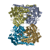
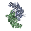
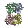
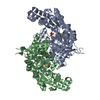
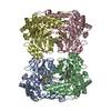
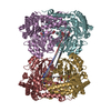
| Title | Co-crystal structure of human serine hydroxymethyltransferase 1 in complex with Pyridoxal 5'-phosphate (PLP) and glycodeoxycholic acid | ||||||
 Components Components | Serine hydroxymethyltransferase, cytosolic | ||||||
 Keywords Keywords | TRANSFERASE / Inhibitor / Complex | ||||||
| Function / homology |  Function and homology information Function and homology informationcellular response to tetrahydrofolate / Carnitine synthesis / carnitine biosynthetic process / purine nucleobase biosynthetic process / serine binding / L-serine catabolic process / glycine metabolic process / L-serine metabolic process / aldehyde-lyase activity / glycine hydroxymethyltransferase ...cellular response to tetrahydrofolate / Carnitine synthesis / carnitine biosynthetic process / purine nucleobase biosynthetic process / serine binding / L-serine catabolic process / glycine metabolic process / L-serine metabolic process / aldehyde-lyase activity / glycine hydroxymethyltransferase / glycine hydroxymethyltransferase activity / glycine biosynthetic process from serine / Metabolism of folate and pterines / tetrahydrofolate metabolic process / tetrahydrofolate interconversion / dTMP biosynthetic process / small molecule binding / folic acid metabolic process / mRNA regulatory element binding translation repressor activity / cellular response to leukemia inhibitory factor / mRNA 5'-UTR binding / pyridoxal phosphate binding / protein homotetramerization / negative regulation of translation / protein homodimerization activity / mitochondrion / extracellular exosome / nucleoplasm / identical protein binding / nucleus / cytosol / cytoplasm Similarity search - Function | ||||||
| Biological species |  Homo sapiens (human) Homo sapiens (human) | ||||||
| Method |  X-RAY DIFFRACTION / SYNCHROTRON / MOLECULAR REPLACEMENT / Resolution: 3.1 Å X-RAY DIFFRACTION / SYNCHROTRON / MOLECULAR REPLACEMENT / Resolution: 3.1 Å | ||||||
 Authors Authors | Ota, T. / Senoo, A. / Ito, S. / Ueno, G. / Nagatoishi, S. / Tsumoto, K. / Sando, S. | ||||||
 Citation Citation | Journal: Iscience / Year: 2021 Title: Structural basis for selective inhibition of human serine hydroxymethyltransferase by secondary bile acid conjugate. Authors: Ota, T. / Senoo, A. / Shirakawa, M. / Nonaka, H. / Saito, Y. / Ito, S. / Ueno, G. / Nagatoishi, S. / Tsumoto, K. / Sando, S. | ||||||
| History |
|
- Structure visualization
| Structure viewer | Molecule: MolmilJmol/JSmol |
|---|
- Downloads & links
-Download
| PDBx/mmCIF format | 6m5w.cif.gz | 108.3 KB | Display | PDBx/mmCIF format |
|---|---|---|---|---|
| PDB format | pdb6m5w.ent.gz | 79.5 KB | Display | PDB format |
| PDBx/mmJSON format | 6m5w.json.gz | Tree view | PDBx/mmJSON format | |
| Others |  Other downloads Other downloads |
-Validation report
| Summary document | 6m5w_validation.pdf.gz | 989.5 KB | Display | wwPDB validaton report |
|---|---|---|---|---|
| Full document | 6m5w_full_validation.pdf.gz | 1011.8 KB | Display | |
| Data in XML | 6m5w_validation.xml.gz | 20.3 KB | Display | |
| Data in CIF | 6m5w_validation.cif.gz | 27 KB | Display | |
| Arichive directory | https://data.pdbj.org/pub/pdb/validation_reports/m5/6m5wftp://data.pdbj.org/pub/pdb/validation_reports/m5/6m5w | HTTPS FTP |
-Related structure data
| Related structure data | 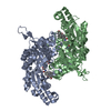 6m5oC 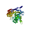 1bj4S S: Starting model for refinement C: citing same article ( |
|---|---|
| Similar structure data |
-Links
 PDBj
PDBj


- Assembly
| Deposited unit | 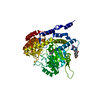
| ||||||||||
|---|---|---|---|---|---|---|---|---|---|---|---|
| 1 | 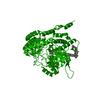
| ||||||||||
| Unit cell |
|
-Components
| #1: Protein | Mass: 55323.867 Da / Num. of mol.: 1 Source method: isolated from a genetically manipulated source Source: (gene. exp.) Homo sapiens (human) / Gene: SHMT1 / Production host:  References: UniProt: P34896, glycine hydroxymethyltransferase |
|---|---|
| #2: Chemical | ChemComp-PLP /   Mass: 247.142 Da / Num. of mol.: 1 / Source method: obtained synthetically / Formula: C8H10NO6P / Feature type: SUBJECT OF INVESTIGATION Mass: 247.142 Da / Num. of mol.: 1 / Source method: obtained synthetically / Formula: C8H10NO6P / Feature type: SUBJECT OF INVESTIGATION |
| #3: Chemical | ChemComp-DXC / (  Mass: 392.572 Da / Num. of mol.: 1 / Source method: obtained synthetically / Formula: C24H40O4 / Feature type: SUBJECT OF INVESTIGATION / Comment: detergent*YM Mass: 392.572 Da / Num. of mol.: 1 / Source method: obtained synthetically / Formula: C24H40O4 / Feature type: SUBJECT OF INVESTIGATION / Comment: detergent*YM |
| #4: Chemical | ChemComp-GLY /   Type: peptide linking / Mass: 75.067 Da / Num. of mol.: 1 / Source method: obtained synthetically / Formula: C2H5NO2 Type: peptide linking / Mass: 75.067 Da / Num. of mol.: 1 / Source method: obtained synthetically / Formula: C2H5NO2 |
| Has ligand of interest | Y |
-Experimental details
-Experiment
| Experiment | Method: X-RAY DIFFRACTION / Number of used crystals: 1 |
|---|
- Sample preparation
| Crystal | Density Matthews: 7.1 Å3/Da / Density % sol: 82.69 % |
|---|---|
| Crystal grow | Temperature: 293 K / Method: vapor diffusion, sitting drop Details: 0.2 M potassium chloride, 0.05 M HEPES pH 7.5, and 35% (v/v) pentaerythritol propoxylate (5/4 PO/OH) |
-Data collection
| Diffraction | Mean temperature: 100 K / Serial crystal experiment: N | ||||||||||||||||||||||||||||||||||||||||||||||||||||||||||||||||||||||||||||||||||||||||||||||||||||
|---|---|---|---|---|---|---|---|---|---|---|---|---|---|---|---|---|---|---|---|---|---|---|---|---|---|---|---|---|---|---|---|---|---|---|---|---|---|---|---|---|---|---|---|---|---|---|---|---|---|---|---|---|---|---|---|---|---|---|---|---|---|---|---|---|---|---|---|---|---|---|---|---|---|---|---|---|---|---|---|---|---|---|---|---|---|---|---|---|---|---|---|---|---|---|---|---|---|---|---|---|---|
| Diffraction source | Source: SYNCHROTRON / Site: SPring-8  / Beamline: BL26B2 / Wavelength: 1 Å / Beamline: BL26B2 / Wavelength: 1 Å | ||||||||||||||||||||||||||||||||||||||||||||||||||||||||||||||||||||||||||||||||||||||||||||||||||||
| Detector | Type: RAYONIX MX225-HS / Detector: CCD / Date: Apr 19, 2019 | ||||||||||||||||||||||||||||||||||||||||||||||||||||||||||||||||||||||||||||||||||||||||||||||||||||
| Radiation | Protocol: SINGLE WAVELENGTH / Monochromatic (M) / Laue (L): M / Scattering type: x-ray | ||||||||||||||||||||||||||||||||||||||||||||||||||||||||||||||||||||||||||||||||||||||||||||||||||||
| Radiation wavelength | Wavelength: 1 Å / Relative weight: 1 | ||||||||||||||||||||||||||||||||||||||||||||||||||||||||||||||||||||||||||||||||||||||||||||||||||||
| Reflection | Resolution: 3.1→46.68 Å / Num. obs: 55751 / % possible obs: 99.9 % / Redundancy: 11.826 % / Biso Wilson estimate: 71.594 Å2 / CC1/2: 0.992 / Rmerge(I) obs: 0.526 / Rrim(I) all: 0.55 / Χ2: 0.726 / Net I/σ(I): 7.31 / Num. measured all: 659305 | ||||||||||||||||||||||||||||||||||||||||||||||||||||||||||||||||||||||||||||||||||||||||||||||||||||
| Reflection shell | Diffraction-ID: 1
|
- Processing
| Software |
| |||||||||||||||||||||||||||||||||||||||||||||||||||||||||||||||
|---|---|---|---|---|---|---|---|---|---|---|---|---|---|---|---|---|---|---|---|---|---|---|---|---|---|---|---|---|---|---|---|---|---|---|---|---|---|---|---|---|---|---|---|---|---|---|---|---|---|---|---|---|---|---|---|---|---|---|---|---|---|---|---|---|
| Refinement | Method to determine structure: MOLECULAR REPLACEMENT Starting model: 1BJ4 Resolution: 3.1→46.68 Å / SU ML: 0.32 / Cross valid method: THROUGHOUT / σ(F): 1.36 / Phase error: 21.15 / Stereochemistry target values: ML
| |||||||||||||||||||||||||||||||||||||||||||||||||||||||||||||||
| Solvent computation | Shrinkage radii: 0.9 Å / VDW probe radii: 1.11 Å / Solvent model: FLAT BULK SOLVENT MODEL | |||||||||||||||||||||||||||||||||||||||||||||||||||||||||||||||
| Displacement parameters | Biso max: 153.76 Å2 / Biso mean: 33.0796 Å2 / Biso min: 6.41 Å2 | |||||||||||||||||||||||||||||||||||||||||||||||||||||||||||||||
| Refinement step | Cycle: final / Resolution: 3.1→46.68 Å
| |||||||||||||||||||||||||||||||||||||||||||||||||||||||||||||||
| LS refinement shell | Refine-ID: X-RAY DIFFRACTION / Rfactor Rfree error: 0 / Total num. of bins used: 8
|
