 Movie
Movie Controller

[English] 日本語
 Yorodumi
- PDB-6cd1: Crystal structure of Medicago truncatula serine hydroxymethyltran... -
+ Open data

- Basic information
| Entry | Database: PDB / ID: 6cd1 | ||||||
|---|---|---|---|---|---|---|---|
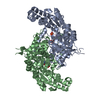
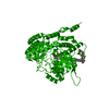
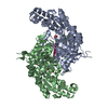
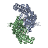
| Title | Crystal structure of Medicago truncatula serine hydroxymethyltransferase 3 (MtSHMT3), complexes with reaction intermediates | ||||||
 Components Components | (Serine hydroxymethyltransferase) x 2 | ||||||
 Keywords Keywords | TRANSFERASE / PLP / one-carbon cycle / tetrahydrofolate / chloroplast / tetramer / PLP-Serine / PLP-Glycine | ||||||
| Function / homology |  Function and homology information Function and homology informationglycine hydroxymethyltransferase / glycine hydroxymethyltransferase activity / : / tetrahydrofolate metabolic process / tetrahydrofolate interconversion / pyridoxal phosphate binding / mitochondrion Similarity search - Function | ||||||
| Biological species |  | ||||||
| Method |  X-RAY DIFFRACTION / SYNCHROTRON / MOLECULAR REPLACEMENT / Resolution: 1.91 Å X-RAY DIFFRACTION / SYNCHROTRON / MOLECULAR REPLACEMENT / Resolution: 1.91 Å | ||||||
 Authors Authors | Ruszkowski, M. / Sekula, B. / Ruszkowska, A. / Dauter, Z. | ||||||
 Citation Citation | Journal: Front Plant Sci / Year: 2018 Title: Chloroplastic Serine Hydroxymethyltransferase FromMedicago truncatula: A Structural Characterization. Authors: Ruszkowski, M. / Sekula, B. / Ruszkowska, A. / Dauter, Z. | ||||||
| History |
|
- Structure visualization
| Structure viewer | Molecule: MolmilJmol/JSmol |
|---|
- Downloads & links
-Download
| PDBx/mmCIF format | 6cd1.cif.gz | 1.4 MB | Display | PDBx/mmCIF format |
|---|---|---|---|---|
| PDB format | pdb6cd1.ent.gz | 1.1 MB | Display | PDB format |
| PDBx/mmJSON format | 6cd1.json.gz | Tree view | PDBx/mmJSON format | |
| Others |  Other downloads Other downloads |
-Validation report
| Arichive directory | https://data.pdbj.org/pub/pdb/validation_reports/cd/6cd1ftp://data.pdbj.org/pub/pdb/validation_reports/cd/6cd1 | HTTPS FTP |
|---|
-Related structure data
| Related structure data |  6cczSC  6cd0C S: Starting model for refinement C: citing same article ( |
|---|---|
| Similar structure data |
-Links
 PDBj
PDBj

- Assembly
| Deposited unit | 
| ||||||||
|---|---|---|---|---|---|---|---|---|---|
| 1 | 
| ||||||||
| 2 | 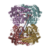
| ||||||||
| Unit cell |
|
-Components
-Protein , 2 types, 8 molecules ABCDFEGH
| #1: Protein | Mass: 49588.469 Da / Num. of mol.: 5 Source method: isolated from a genetically manipulated source Source: (gene. exp.) Production host:  References: UniProt: G7ILW0, glycine hydroxymethyltransferase #2: Protein | Mass: 49816.586 Da / Num. of mol.: 3 Source method: isolated from a genetically manipulated source Source: (gene. exp.) Production host: References: UniProt: G7ILW0, glycine hydroxymethyltransferase |
|---|
-Non-polymers , 6 types, 2049 molecules 


| #3: Chemical |  Mass: 336.235 Da / Num. of mol.: 2 / Source method: obtained synthetically / Formula: C11H17N2O8P / Feature type: SUBJECT OF INVESTIGATION Mass: 336.235 Da / Num. of mol.: 2 / Source method: obtained synthetically / Formula: C11H17N2O8P / Feature type: SUBJECT OF INVESTIGATION#4: Chemical | ChemComp-EDO /  Mass: 62.068 Da / Num. of mol.: 8 / Source method: obtained synthetically / Formula: C2H6O2 Mass: 62.068 Da / Num. of mol.: 8 / Source method: obtained synthetically / Formula: C2H6O2#5: Chemical |  Mass: 306.209 Da / Num. of mol.: 2 / Source method: obtained synthetically / Formula: C10H15N2O7P / Feature type: SUBJECT OF INVESTIGATION Mass: 306.209 Da / Num. of mol.: 2 / Source method: obtained synthetically / Formula: C10H15N2O7P / Feature type: SUBJECT OF INVESTIGATION#6: Chemical |  Mass: 59.044 Da / Num. of mol.: 3 / Source method: obtained synthetically / Formula: C2H3O2 Mass: 59.044 Da / Num. of mol.: 3 / Source method: obtained synthetically / Formula: C2H3O2#7: Chemical |  Type: peptide linking / Mass: 75.067 Da / Num. of mol.: 2 / Source method: obtained synthetically / Formula: C2H5NO2 Type: peptide linking / Mass: 75.067 Da / Num. of mol.: 2 / Source method: obtained synthetically / Formula: C2H5NO2#8: Water | ChemComp-HOH / | Mass: 18.015 Da / Num. of mol.: 2032 / Source method: isolated from a natural source / Formula: H2O |
|---|
-Experimental details
-Experiment
| Experiment | Method: X-RAY DIFFRACTION / Number of used crystals: 1 |
|---|
- Sample preparation
| Crystal | Density Matthews: 2.19 Å3/Da / Density % sol: 43.96 % |
|---|---|
| Crystal grow | Temperature: 292 K / Method: vapor diffusion, hanging drop / pH: 6.5 Details: 75 mM MES pH 6.5, 19% PEG3350 and 150 mM ammonium acetate. The mature crystals were soaked with 200 mM Ser for 2 h and cryoprotected by the addition of ethylene glycol to a final concentration of 20% |
-Data collection
| Diffraction | Mean temperature: 100 K |
|---|---|
| Diffraction source | Source: SYNCHROTRON / Site: APS  / Beamline: 19-ID / Wavelength: 0.9792 Å / Beamline: 19-ID / Wavelength: 0.9792 Å |
| Detector | Type: DECTRIS PILATUS 6M / Detector: PIXEL / Date: Nov 15, 2017 |
| Radiation | Protocol: SINGLE WAVELENGTH / Monochromatic (M) / Laue (L): M / Scattering type: x-ray |
| Radiation wavelength | Wavelength: 0.9792 Å / Relative weight: 1 |
| Reflection | Resolution: 1.91→50 Å / Num. obs: 263165 / % possible obs: 98.6 % / Redundancy: 4.7 % / Rmerge(I) obs: 0.063 / Rrim(I) all: 0.071 / Net I/σ(I): 13.3 |
| Reflection shell | Resolution: 1.91→2.02 Å / Redundancy: 4.7 % / Rmerge(I) obs: 0.73 / Mean I/σ(I) obs: 1.9 / Num. unique obs: 41826 / Rrim(I) all: 0.83 / % possible all: 97.3 |
- Processing
| Software |
| ||||||||||||||||||||||||||||||||||||||||||||||||||||||||||||||||||||||||||||||||||||||||||||||||||||||||||||||||||||||||||||||||||||||||||||||||||||||||||||||||||||||||||||||||||||||
|---|---|---|---|---|---|---|---|---|---|---|---|---|---|---|---|---|---|---|---|---|---|---|---|---|---|---|---|---|---|---|---|---|---|---|---|---|---|---|---|---|---|---|---|---|---|---|---|---|---|---|---|---|---|---|---|---|---|---|---|---|---|---|---|---|---|---|---|---|---|---|---|---|---|---|---|---|---|---|---|---|---|---|---|---|---|---|---|---|---|---|---|---|---|---|---|---|---|---|---|---|---|---|---|---|---|---|---|---|---|---|---|---|---|---|---|---|---|---|---|---|---|---|---|---|---|---|---|---|---|---|---|---|---|---|---|---|---|---|---|---|---|---|---|---|---|---|---|---|---|---|---|---|---|---|---|---|---|---|---|---|---|---|---|---|---|---|---|---|---|---|---|---|---|---|---|---|---|---|---|---|---|---|---|
| Refinement | Method to determine structure: MOLECULAR REPLACEMENT Starting model: 6ccz Resolution: 1.91→46.65 Å / Cor.coef. Fo:Fc: 0.965 / Cor.coef. Fo:Fc free: 0.948 / SU B: 10.953 / SU ML: 0.154 / Cross valid method: THROUGHOUT / ESU R: 0.168 / ESU R Free: 0.153 / Details: HYDROGENS HAVE BEEN ADDED IN THE RIDING POSITIONS
| ||||||||||||||||||||||||||||||||||||||||||||||||||||||||||||||||||||||||||||||||||||||||||||||||||||||||||||||||||||||||||||||||||||||||||||||||||||||||||||||||||||||||||||||||||||||
| Solvent computation | Ion probe radii: 0.8 Å / Shrinkage radii: 0.8 Å / VDW probe radii: 1.2 Å | ||||||||||||||||||||||||||||||||||||||||||||||||||||||||||||||||||||||||||||||||||||||||||||||||||||||||||||||||||||||||||||||||||||||||||||||||||||||||||||||||||||||||||||||||||||||
| Displacement parameters | Biso mean: 40.906 Å2
| ||||||||||||||||||||||||||||||||||||||||||||||||||||||||||||||||||||||||||||||||||||||||||||||||||||||||||||||||||||||||||||||||||||||||||||||||||||||||||||||||||||||||||||||||||||||
| Refinement step | Cycle: 1 / Resolution: 1.91→46.65 Å
| ||||||||||||||||||||||||||||||||||||||||||||||||||||||||||||||||||||||||||||||||||||||||||||||||||||||||||||||||||||||||||||||||||||||||||||||||||||||||||||||||||||||||||||||||||||||
| Refine LS restraints |
|