 Movie
Movie Controller

[English] 日本語
 Yorodumi
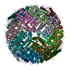
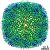
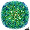
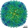
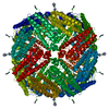
- PDB-6m54: Human apo ferritin frozen on TEM grid with Amorphous nickel titan... -
+ Open data

- Basic information
| Entry | Database: PDB / ID: 6m54 | |||||||||||||||
|---|---|---|---|---|---|---|---|---|---|---|---|---|---|---|---|---|
| Title | Human apo ferritin frozen on TEM grid with Amorphous nickel titanium alloy supporting film | |||||||||||||||
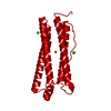
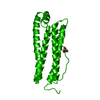
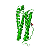
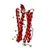
 Components Components | Ferritin heavy chain | |||||||||||||||
 Keywords Keywords | OXIDOREDUCTASE / Apoferritin heavy chain / Homo sapiens / METAL TRANSPORT | |||||||||||||||
| Function / homology |  Function and homology information Function and homology informationiron ion sequestering activity / ferritin complex / Scavenging by Class A Receptors / Golgi Associated Vesicle Biogenesis / ferroxidase / negative regulation of ferroptosis / autolysosome / ferroxidase activity / negative regulation of fibroblast proliferation / ferric iron binding ...iron ion sequestering activity / ferritin complex / Scavenging by Class A Receptors / Golgi Associated Vesicle Biogenesis / ferroxidase / negative regulation of ferroptosis / autolysosome / ferroxidase activity / negative regulation of fibroblast proliferation / ferric iron binding / autophagosome / iron ion transport / ferrous iron binding / Iron uptake and transport / tertiary granule lumen / ficolin-1-rich granule lumen / intracellular iron ion homeostasis / immune response / iron ion binding / negative regulation of cell population proliferation / Neutrophil degranulation / extracellular exosome / extracellular region / identical protein binding / nucleus / cytoplasm / cytosol Similarity search - Function | |||||||||||||||
| Biological species |  Homo sapiens (human) Homo sapiens (human) | |||||||||||||||
| Method | ELECTRON MICROSCOPY / single particle reconstruction / cryo EM / Resolution: 2.4 Å | |||||||||||||||
 Authors Authors | Huang, X. / Zhang, L. / Wen, Z. / Chen, H. / Li, S. / Ji, G. / Yin, C. / Sun, F. | |||||||||||||||
| Funding support |  China, 4items China, 4items
| |||||||||||||||
 Citation Citation | Journal: Prog Biophys Mol Biol / Year: 2020 Title: Amorphous nickel titanium alloy film: A new choice for cryo electron microscopy sample preparation. Authors: Xiaojun Huang / Lei Zhang / Zuoling Wen / Hui Chen / Shuoguo Li / Gang Ji / Chang-Cheng Yin / Fei Sun / Abstract: Cryo-electron microscopy (cryoEM) has become one of the most important approach for structural biology. However, barriers are still there for an increased successful rate, a better resolution and ...Cryo-electron microscopy (cryoEM) has become one of the most important approach for structural biology. However, barriers are still there for an increased successful rate, a better resolution and improved efficiency from sample preparation, data collection to image processing. CryoEM sample preparation is one of the bottlenecks with many efforts made recently, including the optimization of supporting substrate (e.g. ultra-thin carbon, graphene, pure gold, 2d crystal of streptavidin, and affinity modification), which was aimed to solve air-water interface problem, or reduce beam induced motion (BIM), or change particle distribution in the grid hole. Here, we report another effort of developing a new supporting substrate, the amorphous nickel-titanium alloy (ANTA) film, for cryoEM sample preparation as a layer of holey supporting film covering on TEM grid. Our investigations showed advantages of ANTA film in comparison with conventional carbon film, including much better electron conductivity and trace non-specific interaction with protein. These advantages yield less BIM and significantly improved particle distribution during cryoEM experiment of human apo-ferritn, thus resulting an improved reconstruction resolution from a reduced number of micrographs and particles. Unlike the pure gold film, the usage of the ANTA film is just same with the carbon film, compatible to conventional automatic cryoEM data collection procedure. | |||||||||||||||
| History |
|
- Structure visualization
| Movie |
Movie viewer |
|---|---|
| Structure viewer | Molecule: MolmilJmol/JSmol |
- Downloads & links
-Download
| PDBx/mmCIF format | 6m54.cif.gz | 835.8 KB | Display | PDBx/mmCIF format |
|---|---|---|---|---|
| PDB format | pdb6m54.ent.gz | 591.5 KB | Display | PDB format |
| PDBx/mmJSON format | 6m54.json.gz | Tree view | PDBx/mmJSON format | |
| Others |  Other downloads Other downloads |
-Validation report
| Arichive directory | https://data.pdbj.org/pub/pdb/validation_reports/m5/6m54ftp://data.pdbj.org/pub/pdb/validation_reports/m5/6m54 | HTTPS FTP |
|---|
-Related structure data
| Related structure data |  30084MC  6m52C C: citing same article ( M: map data used to model this data |
|---|---|
| Similar structure data | |
| EM raw data | EMPIAR-10386 (Title: Human apo ferritin frozen on TEM grid with Amorphous nickel titanium alloy supporting film Data size: 134.5 Data #1: Human apo ferritin frozen on TEM grid with Amorphous nickel titanium alloy supporting film [micrographs - multiframe]) |


-Links
 PDBj
PDBj


- Assembly
| Deposited unit | 
|
|---|---|
| 1 |
|
-Components
| #1: Protein | Mass: 21255.656 Da / Num. of mol.: 24 Source method: isolated from a genetically manipulated source Source: (gene. exp.) Homo sapiens (human) / Gene: FTH1, FTH, FTHL6, OK/SW-cl.84, PIG15 / Production host:  #2: Chemical | ChemComp-FE2 /   Mass: 55.845 Da / Num. of mol.: 6 / Source method: obtained synthetically / Formula: Fe Mass: 55.845 Da / Num. of mol.: 6 / Source method: obtained synthetically / Formula: Fe#3: Water | ChemComp-HOH / |  Mass: 18.015 Da / Num. of mol.: 8 / Source method: isolated from a natural source / Formula: H2O Mass: 18.015 Da / Num. of mol.: 8 / Source method: isolated from a natural source / Formula: H2OHas ligand of interest | N | |
|---|
-Experimental details
-Experiment
| Experiment | Method: ELECTRON MICROSCOPY |
|---|---|
| EM experiment | Aggregation state: PARTICLE / 3D reconstruction method: single particle reconstruction |
- Sample preparation
| Component | Name: apoferritin heavy chain, 24 homologous polymer / Type: COMPLEX Details: frozen on TEM grid with amorphous nickel titanium alloy supporting film Entity ID: #1 / Source: RECOMBINANT | |||||||||||||||
|---|---|---|---|---|---|---|---|---|---|---|---|---|---|---|---|---|
| Molecular weight | Value: 0.44 MDa / Experimental value: NO | |||||||||||||||
| Source (natural) | Organism: Homo sapiens (human) | |||||||||||||||
| Source (recombinant) | Organism: | |||||||||||||||
| Buffer solution | pH: 8 | |||||||||||||||
| Buffer component |
| |||||||||||||||
| Specimen | Conc.: 1.3 mg/ml / Embedding applied: NO / Shadowing applied: NO / Staining applied: NO / Vitrification applied: YES | |||||||||||||||
| Specimen support | Grid material: GOLD / Grid mesh size: 300 divisions/in. / Grid type: Homemade | |||||||||||||||
| Vitrification | Instrument: LEICA EM GP / Cryogen name: ETHANE |
- Electron microscopy imaging
| Experimental equipment |  Model: Titan Krios / Image courtesy: FEI Company |
|---|---|
| Microscopy | Model: FEI TITAN KRIOS |
| Electron gun | Electron source:  FIELD EMISSION GUN / Accelerating voltage: 300 kV / Illumination mode: FLOOD BEAM FIELD EMISSION GUN / Accelerating voltage: 300 kV / Illumination mode: FLOOD BEAM |
| Electron lens | Mode: BRIGHT FIELD / Cs: 2.7 mm |
| Image recording | Electron dose: 60 e/Å2 / Detector mode: SUPER-RESOLUTION / Film or detector model: GATAN K2 SUMMIT (4k x 4k) |
- Processing
| Software | Name: PHENIX / Version: 1.16_3549 / Classification: refinement / Contact author: Paul D. Adams / Contact author email: pdadams[at]lbl.gov / Language: Python/C++ / URL: https://www.phenix-online.org/ / Type: program | ||||||||||||||||||||||||
|---|---|---|---|---|---|---|---|---|---|---|---|---|---|---|---|---|---|---|---|---|---|---|---|---|---|
| EM software | Name: SerialEM / Version: 3.6 / Category: image acquisition | ||||||||||||||||||||||||
| CTF correction | Type: PHASE FLIPPING ONLY | ||||||||||||||||||||||||
| Symmetry | Point symmetry: O (octahedral) | ||||||||||||||||||||||||
| 3D reconstruction | Resolution: 2.4 Å / Resolution method: FSC 0.143 CUT-OFF / Num. of particles: 91537 / Symmetry type: POINT | ||||||||||||||||||||||||
| Refinement | Cross valid method: NONE Stereochemistry target values: GeoStd + Monomer Library + CDL v1.2 | ||||||||||||||||||||||||
| Displacement parameters | Biso mean: 68.05 Å2 | ||||||||||||||||||||||||
| Refine LS restraints |
|
