 Movie
Movie Controller


+ Open data

- Basic information
| Entry | Database: PDB / ID: 1rcc | |||||||||
|---|---|---|---|---|---|---|---|---|---|---|
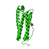
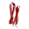
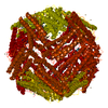
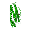
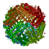
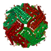
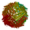
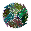
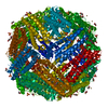
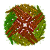
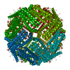
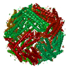
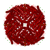
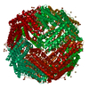
| Title | BULLFROG RED CELL L FERRITIN TARTRATE/MG/PH 5.5 | |||||||||
 Components Components | L FERRITIN | |||||||||
 Keywords Keywords | IRON STORAGE | |||||||||
| Function / homology |  Function and homology information Function and homology informationferric iron binding / iron ion transport / ferrous iron binding / intracellular iron ion homeostasis / cytoplasm Similarity search - Function | |||||||||
| Biological species | Rana catesbeiana (American bullfrog) | |||||||||
| Method |  X-RAY DIFFRACTION / Resolution: 2.4 Å X-RAY DIFFRACTION / Resolution: 2.4 Å | |||||||||
 Authors Authors | Trikha, J. / Theil, E.C. / Allewell, N.M. | |||||||||
 Citation Citation | Journal: J.Mol.Biol. / Year: 1995 Title: High resolution crystal structures of amphibian red-cell L ferritin: potential roles for structural plasticity and solvation in function. Authors: Trikha, J. / Theil, E.C. / Allewell, N.M. #1: Journal: Proteins / Year: 1994Title: Crystallization and Structural Analysis of Bullfrog Red Cell L-Subunit Ferritins Authors: Trikha, J. / Waldo, G.S. / Lewandowski, F.A. / Ha, Y. / Theil, E.C. / Weber, P.C. / Allewell, N.M. | |||||||||
| History |
|
- Structure visualization
| Structure viewer | Molecule: MolmilJmol/JSmol |
|---|
- Downloads & links
-Download
| PDBx/mmCIF format | 1rcc.cif.gz | 47.4 KB | Display | PDBx/mmCIF format |
|---|---|---|---|---|
| PDB format | pdb1rcc.ent.gz | 34.8 KB | Display | PDB format |
| PDBx/mmJSON format | 1rcc.json.gz | Tree view | PDBx/mmJSON format | |
| Others |  Other downloads Other downloads |
-Validation report
| Arichive directory | https://data.pdbj.org/pub/pdb/validation_reports/rc/1rccftp://data.pdbj.org/pub/pdb/validation_reports/rc/1rcc | HTTPS FTP |
|---|
-Related structure data


-Links
 PDBj
PDBj


- Assembly
| Deposited unit | 
| ||||||||
|---|---|---|---|---|---|---|---|---|---|
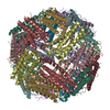
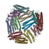
| 1 | x 24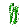
| ||||||||
| Unit cell |
| ||||||||
| Atom site foot note | 1: CIS PROLINE - PRO 132 |
-Components
| #1: Protein | Mass: 19735.186 Da / Num. of mol.: 1 / Mutation: E57A, E58A, E59A, E61A Source method: isolated from a genetically manipulated source Source: (gene. exp.) Rana catesbeiana (American bullfrog) / Cell: RED CELL / Gene: CDNA / Plasmid: PET 3A / Gene (production host): CDNA / Production host:  |
|---|---|
| #2: Chemical | ChemComp-BET /   Mass: 118.154 Da / Num. of mol.: 1 / Source method: obtained synthetically / Formula: C5H12NO2 Mass: 118.154 Da / Num. of mol.: 1 / Source method: obtained synthetically / Formula: C5H12NO2 |
| #3: Water | ChemComp-HOH /  Mass: 18.015 Da / Num. of mol.: 29 / Source method: isolated from a natural source / Formula: H2O Mass: 18.015 Da / Num. of mol.: 29 / Source method: isolated from a natural source / Formula: H2O |
-Experimental details
-Experiment
| Experiment | Method: X-RAY DIFFRACTION / Number of used crystals: 1 |
|---|
- Sample preparation
| Crystal | Density Matthews: 3.17 Å3/Da / Density % sol: 61.23 % | ||||||||||||||||||||||||
|---|---|---|---|---|---|---|---|---|---|---|---|---|---|---|---|---|---|---|---|---|---|---|---|---|---|
| Crystal grow | pH: 5.5 / Details: pH 5.5 | ||||||||||||||||||||||||
| Crystal | *PLUS Density % sol: 54 % | ||||||||||||||||||||||||
| Crystal grow | *PLUS Method: unknown | ||||||||||||||||||||||||
| Components of the solutions | *PLUS
|
-Data collection
| Diffraction source | Wavelength: 1.5418 Å |
|---|---|
| Detector | Type: NICOLET / Detector: AREA DETECTOR / Date: Jun 15, 1992 |
| Radiation | Monochromatic (M) / Laue (L): M / Scattering type: x-ray |
| Radiation wavelength | Wavelength: 1.5418 Å / Relative weight: 1 |
| Reflection | Resolution: 2.4→10 Å / Num. obs: 13375 / % possible obs: 95.5 % / Observed criterion σ(I): 2 / Redundancy: 2 % / Rmerge(I) obs: 0.139 |
| Reflection | *PLUS Rmerge(I) obs: 0.139 |
- Processing
| Software |
| ||||||||||||||||||||||||||||||||||||||||||||||||||||||||||||
|---|---|---|---|---|---|---|---|---|---|---|---|---|---|---|---|---|---|---|---|---|---|---|---|---|---|---|---|---|---|---|---|---|---|---|---|---|---|---|---|---|---|---|---|---|---|---|---|---|---|---|---|---|---|---|---|---|---|---|---|---|---|
| Refinement | Resolution: 2.4→10 Å / σ(F): 2
| ||||||||||||||||||||||||||||||||||||||||||||||||||||||||||||
| Displacement parameters | Biso mean: 11 Å2 | ||||||||||||||||||||||||||||||||||||||||||||||||||||||||||||
| Refine analyze | Luzzati coordinate error obs: 0.15 Å | ||||||||||||||||||||||||||||||||||||||||||||||||||||||||||||
| Refinement step | Cycle: LAST / Resolution: 2.4→10 Å
| ||||||||||||||||||||||||||||||||||||||||||||||||||||||||||||
| Refine LS restraints |
| ||||||||||||||||||||||||||||||||||||||||||||||||||||||||||||
| Software | *PLUS Name: X-PLOR / Classification: refinement | ||||||||||||||||||||||||||||||||||||||||||||||||||||||||||||
| Refinement | *PLUS | ||||||||||||||||||||||||||||||||||||||||||||||||||||||||||||
| Solvent computation | *PLUS | ||||||||||||||||||||||||||||||||||||||||||||||||||||||||||||
| Displacement parameters | *PLUS |
